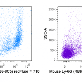
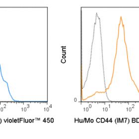
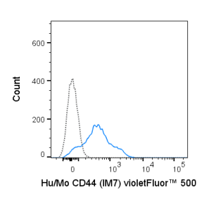
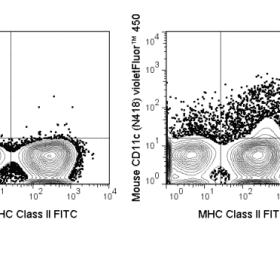
The M1/70 antibody reacts with human and mouse CD11b, also known as integrin αalpha M. This 165-170 kDa cell surface glycoprotein is part of a family of integrin αreceptors that mediate adhesion between ≥ ≥ ≥ cells (cell-cell) and components of the extracellular matrix, e.g. fibrinogen (cell-matrix). In addition, integrin αs are active signaling receptors which recruit leukocytes to inflammatory sites and promote cell activation. Complete, functional integrin αreceptors consist of distinct combinations of integrin αchains which are differentially expressed. integrin αalpha M (CD11b) assembles with integrin αbeta-2 (CD18) into a receptor known as Macrophage Antigen-1 (Mac-1) or complement receptor type 3 (CR3). This receptor binds and induces intracellular signaling through ICAM-1 on endothelial cells and can also facilitate removal of iC3b bearing foreign cells.
The M1/70 antibody is widely used as a marker for CD11b expression on mouse macrophages, granulocytes, neutrophils, and NK cells. The antibody is also reported to be cross-reactive for Rhesus macaque CD11b.
Product Details
| Name | violetFluor™ 450 Anti-Human/Mouse CD11b (M1/70) |
|---|---|
| Cat. No. | 75-0112 |
| Alternative Names | CD11, Mac-1, integrin α?M, CR3, ITGAM |
| Gene ID | 16409 / 3684 |
| Clone | M1/70 |
| Isotype | Rat IgG2b, κ |
| Reactivity | Human, Mouse |
| Cross Reactivity | Chimpanzee, Baboon, Cynomolgus, Rhesus |
| Format | violetFluor™ 450 |
| Application | Flow Cytometry |
| Citations* | Lefort CT, Rossaint J, Moser M, Petrich BG, Zarbock A, Monkley SJ, Critchley DR, Ginsberg MH, Fassler R, and Ley K. 2012. Blood. 119:4275-4282. (in vitro blocking)
Grewal JS, Pilgrim MJ, Grewal S, Kasman L, Werner P, Bruorton ME, London SD, and London L. 2011. FASEB J. 25:1680-1696. (Immunofluorescence microscopy – frozen tissue) Kim W-K, Sun Y, Do H, Autissier P, Halpern EF, Piatak M, Lifson JD, Burdo TH, McGrath MS, and Williams K. 2010. J. Leukoc. Biol. 87: 557-567. (Flow Cytometry – Rhesus macaque) Roland CL, Dineen SP, Lynn KD, Sullivan LA, Dellinger MT, Sadegh L, Sullivan JP, Shames DS, and Brekken RA. 2009. Mol. Cancer Ther. 8:1761-1771. (Immunofluorescence microscopy – frozen tissue) Sorg H, Lorch B, Jaster R, Fitzner B, Ibrahim S, Holzhueter S, Nizze H, and Vollmar B. 2008. Am. J. Physio. Gastrointest. Liver Physiol. 295: G1274-1280. (Immunohistochemistry – formalin-fixed paraffin embedded tissue) Kim DD, Miwa T, Kimura Y, Schwendener RA, van Lookeren Campagne M, and Song W-C. 2008. Blood. 112:1109-1119. (in vivo blocking) Ou R, Zhang M, Huang L, Flavell RA, Koni PA, and Moskophidis D. 2008. J. Virol. 82:2952-2965. (Immunohistochemistry – OCT embedded frozen tissue) Nutt SL, Metcalf D, D’Amico A, Polli M, and Wu L. 2005. J. Exp. Med. 201:221-231. (Immunomagnetic bead depletion) Whiteland JL, Nicholls SM, Shimeld C, Easty DL, Williams NA, and Hill TJ. 1995. J. Histochem. Cytochem. 43:313-320. (Immunohistochemistry – frozen tissue, paraffin embedded tissue) Miller LJ, Schwarting R, and Springer TA. 1986. J. Immunol. 137:2891-2900. (Immunoprecipitation) |
Application Key:FC = Flow Cytometry; FA = Functional Assays; ELISA = Enzyme-Linked Immunosorbent Assay; ICC = Immunocytochemistry; IF = Immunofluorescence Microscopy; IHC = Immunohistochemistry; IHC-F = Immunohistochemistry, Frozen Tissue; IHC-P = Immunohistochemistry, Paraffin-Embedded Tissue; IP = Immunoprecipitation; WB = Western Blot; EM = Electron Microscopy
*Tonbo Biosciences tests all antibodies by flow cytometry. Citations are provided as a resource for additional applications that have not been validated by Tonbo Biosciences. Please choose the appropriate format for each application and consult the Materials and Methods section for additional details about the use of any product in these publications.


