| Cat No. | Size | Price |
|---|---|---|
| 79-1619-U025 | 25 µg | $88.00 |
| 79-1619-U100 | 100 µg | $220.00 |
Description
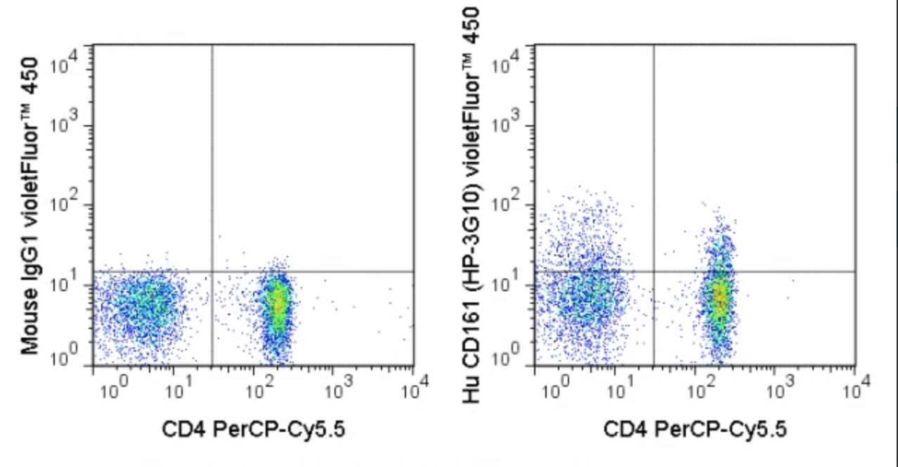
The HP-3G10 antibody is specific for human CD161, also known as NKR-P1A, a type II transmembrane lectin-like receptor and member of the killer cell lectin-like receptor (KLR) family. CD161 exists as a homodimer which is prominently expressed on natural killer (NK) and NKT cells, where it is proposed to regulate the function of both cell types. CD161 is also found on T cell subsets, including T regulatory cells (Tregs), memory/effector CD4+ T cells, and CD8+ T cells. Th17 cells have been demonstrated to co-express CD161, as surface IL-17A+ cells are contained within the CD161+ fraction of CD4 T cells, so that CD161 (in combination with CCR6) is often used as a marker for enrichment of Th17 cells.
The HP-3G10 antibody may be used for flow cytometric analysis of CD161 on NK and NKT cells, as well as on various T cell subsets. The antibody is also reported to be cross-reactive with Baboon, Chimpanzee and Rhesus CD161.
Product Details
| Name | violetFluor™ 450 Anti-Human CD161 (HP-3G10) |
|---|---|
| Cat. No. | 75-1619 |
| Alternative Names | NKR-P1A, NKRP1A |
| Gene ID | 3820 |
| Clone | HP-3G10 |
| Isotype | Mouse IgG1, kappa |
| Reactivity | Human |
| Cross Reactivity | Chimpanzee, Baboon, Rhesus |
| Format | violetFluor™ 450 |
| Application | Flow Cytometry |
| Citations* | Yamada H, Nakashima Y, Okazaki K, Mawatari T, Fukushi J-I, Oyamada A, Fujimura K, Iwamoto Y, and Yoshikai Y. 2011. J. Rheumatol. 38: 1569-1575. (Flow cytometry)
Fogal B, Yi T, Wang C, Rao DA, Lebastchi A, Kulkarni S, Tellides G, and Pober JS. 2011. J. Immunol. 187: 6268-6280. (in vitro depletion) Pozo, D, Vales-Gomez, Mavaddat N, Williamson SC, Chisholm SE, and Reyburn H. 2006. J. Immunol. 176: 2397-2406. (Western blot) Exley M, Porcelli S, Furman M, Garcia J, and Balk S. 1998. J. Exp. Med. 188: 867-876. (in vitro blocking, Western blot) |
Application Key:FC = Flow Cytometry; FA = Functional Assays; ELISA = Enzyme-Linked Immunosorbent Assay; ICC = Immunocytochemistry; IF = Immunofluorescence Microscopy; IHC = Immunohistochemistry; IHC-F = Immunohistochemistry, Frozen Tissue; IHC-P = Immunohistochemistry, Paraffin-Embedded Tissue; IP = Immunoprecipitation; WB = Western Blot; EM = Electron Microscopy
*Tonbo Biosciences tests all antibodies by flow cytometry. Citations are provided as a resource for additional applications that have not been validated by Tonbo Biosciences. Please choose the appropriate format for each application and consult the Materials and Methods section for additional details about the use of any product in these publications.
[accordions]
[accordion title=”Protocols”]Technical Date Sheet[/accordion]

