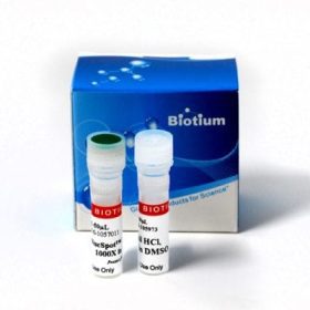
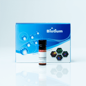
Biotium products are distributed only in Singapore and Thailand.
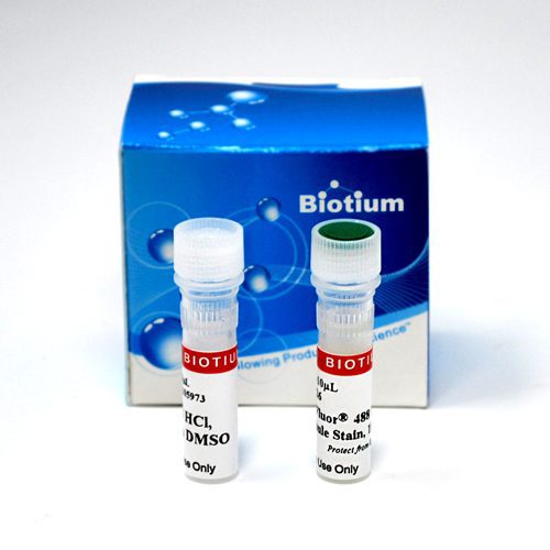
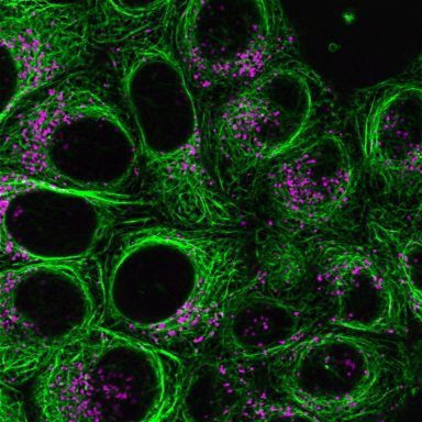
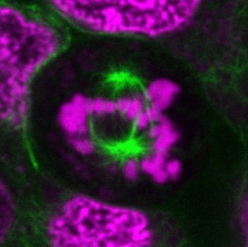
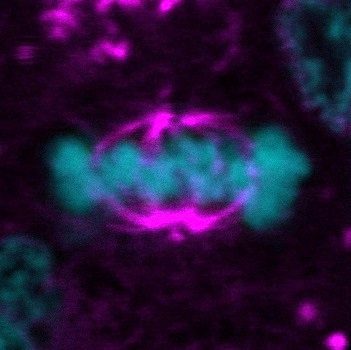
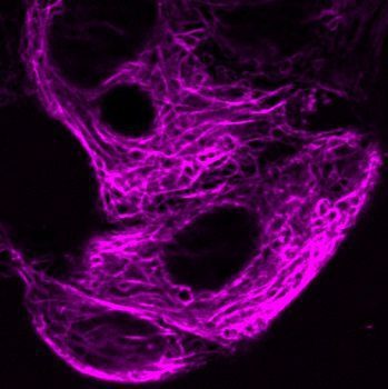
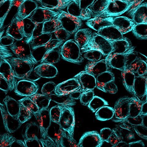
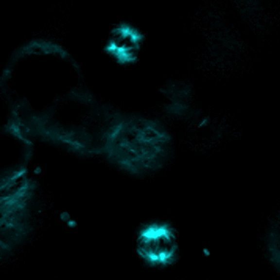
ViaFluor® Live Cell Microtubule Stains are cell-permeable taxol probes for imaging the microtubule cytoskeleton in live cells. Available with blue, green, or far-red fluorescence.
PRODUCT ATTRIBUTES
| Cellular localization | Cytoskeleton, Microtubules |
|---|---|
| Dye | ViaFluor® 405, ViaFluor® 488, ViaFluor® 647 |
| For live or fixed cells | For live/intact cells |
| Assay type/options | Long term staining (24-72h) |
| Cell permeability | Membrane permeant |
| Colors | Blue, Green, Far-red |
PRODUCT DESCRIPTION
Intracellular lipid droplets are cytoplasmic organelles involved in the storage and regulation of triglycerides and cholesterol esters. LipidSpot™ dyes are fluorogenic neutral lipid stains that rapidly accumulate in lipid droplets, where they become brightly fluorescent. The dyes can be used to stain lipid droplets in both live and fixed cells, with no wash step required. Cells also can be fixed and permeabilized after staining. LipidSpot™ stains show minimal background staining of cellular membranes or other organelles, unlike traditional dyes like Nile Red.
LipidSpot™ 488 has excitation around 430 nm, and can be excited equally well at 405 nm or 488 nm. In cells, it stains lipid droplets with bright green fluorescence detectable in the FITC channel. LipidSpot™ 488 has been validated in super-resolution imaging by SIM (Ref. 3).
LipidSpot™ 610 has excitation/emission at ~592/638 nm in cells; it is optimally detected in the Texas Red® channel, but is also bright in the Cy®3 and far-red Cy®5 channels. Therefore, we don’t recommend pairing LipidSpot™ 610 with other red or far-red probes.
Features
- Rapidly and specifically stain lipid droplets
- Stain live or fixed cells, or fix and permeabilize after staining
- Available with green or red/far-red fluorescence
- Supplied as 1000X stock solutions in DMSO
LipidSpot™ 488 stains intracellular membranes in yeast, but LipidSpot™ 610 does not. Both LipidSpot™ dyes can stain gram-positive bacteria, but not gram-negative bacteria. See our Cellular Stains Table for more information on how our dyes stain various organisms.
LipidSpot™ Lipid Droplet Stains
ViaFluor® Live Cell Microtubule Stains are cell-permeable TAXOL® (paclitaxel) probes for imaging the microtubule cytoskeleton in live cells. They are simple, rapid and sensitive stains that can be imaged without a wash step.
TAXOL® binds to polymerized tubulin and stabilizes microtubules, resulting in inhibition of mitosis. However, fluorescent TAXOL® compounds like ViaFluor® Microtubule Stains are less disruptive of microtubule dynamics and cell division, presumably due to lower binding affinity of the fluorescent probe compared to TAXOL® itself. The probes can be used to stain cells for as long as 48 hours to several days. However, the probes may show cytotoxicity and cell cycle arrest with longer incubation times. ViaFluor® Live Cell Microtubule Stains cannot be fixed after staining, and cannot be used with fixed cells or tissues.
ViaFluor® Microtubule Stains are supplied as a 1000X stock solutions in DMSO. They come with a vial of 100 mM verapamil, an efflux pump inhibitor that may improve probe retention and staining in some cell types. Note that this is a higher concentration than previously supplied with the stains, to allow for the use of higher verapamil concentrations (10-100 uM). The optimal concentration of verapamil should be determined by the user for their cell type.
ViaFluor® Live Cell Microtubule Stains are available in three colors:
| Catalog number | Name | Ex/Em | Features |
|---|---|---|---|
| 70064 | ViaFluor® 405 Live Cell Microtubule Stain | 408/452 nm | Blue fluorescence for the DAPI channel |
| 70062 | ViaFluor® 488 Live Cell Microtubule Stain | 500/515 nm | Green fluorescence for the FITC channel |
| 70063 | ViaFluor® 647 Live Cell Microtubule Stain | 650/675 nm | Far-red fluorescence for the Cy®5 channel; compatible with SIM or STED |
Learn more about our other fluorescent cellular stains for nucleus, mitochondria, lysosomes, and more.
Documents
Protocols
SDS
Supporting Documents

