| Cat No. | Size | Price |
|---|---|---|
| 60-0564-T025 | 25 tests | $71.00 |
| 60-0564-T100 | 100 tests | $159.00 |
Description
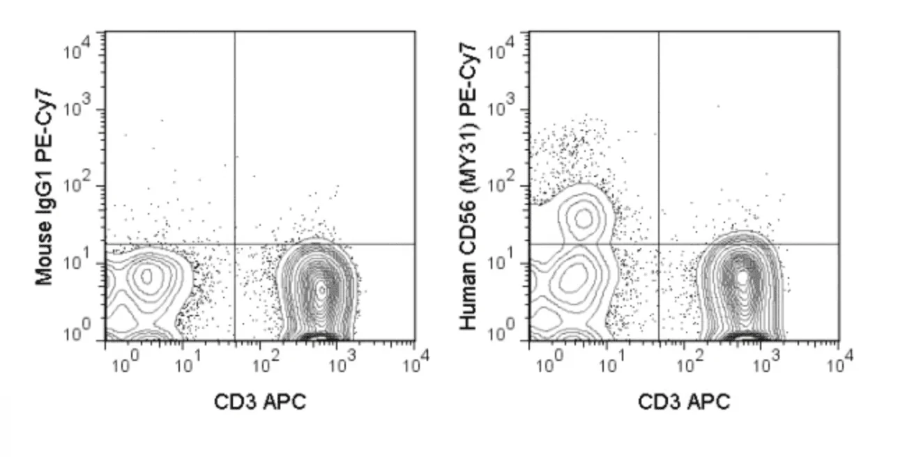
The MY31 antibody reacts with human CD56, also known as the neural cell adhesion molecule (NCAM), a glycoprotein which is a member of the immunoglobulin superfamily. The 140 kDa isoform of CD56 is expressed on human NK cells and NK-T cells, with increased expression levels on activated NK lymphocytes. The CD56 antigen is also expressed by neurons and is reported to play a role in nervous system development and neural cell-to-cell adhesion.
Clone MY31 also reacts with a subset of CD14+ monocytes in non-human primates, and is reported to be cross-reactive with Chimpanzee, Cynomolgus and Rhesus.
Product Details
| Name | PE-Cyanine7 Anti-Human CD56 (NCAM) (MY31) |
|---|---|
| Cat. No. | 60-0564 |
| Alternative Names | neural cell adhesion molecule (NCAM), Leu-19 |
| Gene ID | 4684 |
| Clone | MY31 |
| Isotype | Mouse IgG1, kappa |
| Reactivity | Human |
| Cross Reactivity | Chimpanzee, Cynomolgus, Rhesus |
| Format | PE-Cyanine7 |
| Application | Flow Cytometry |
| Citations* | Lanier LL, Testi R, Bindl J and Phillips JH. 1989. J Exp Med. 169: 2233-2238.
Schlossman SF, Boumsell L, Gilks W et al., eds. 1995. Leucocyte Typing V: White Cell Differentiation Antigens. Oxford University Press. Carter DL, Shieh TM, Blosser RL, Chadwick KR, Margolick JB, Hidreth JE, Clements JE and Zink MC. 1999. Cytometry. 37(1): 41-50. Chan WK, Suwannasaen D, Throm RE, Li Y, Eldridge PW, Houston J, Gray JT, Pui C-H and Leung W. 2015. Leukemia. 29: 387-395. (Flow cytometry) Woltman AM, Op den Brouw ML, Biesta PJ, Shi CC and Janssen HLA. 2011. PLoS ONE 6(1): e15324. doi: 10.1371/journal.pone.0015324. (Flow cytometry) Bleul CC, Wu L, Hoxie JA, Springer TA and Mackay CR. 1997. Proc Natl Acad Sci USA. 94(5): 1925-1930. (Flow cytometry) Brown K and Barratt-Boyes SM. 2009. J Med Primatol. 38(4): 272-278. (Flow cytometry – Rhesus) |
Application Key:FC = Flow Cytometry; FA = Functional Assays; ELISA = Enzyme-Linked Immunosorbent Assay; ICC = Immunocytochemistry; IF = Immunofluorescence Microscopy; IHC = Immunohistochemistry; IHC-F = Immunohistochemistry, Frozen Tissue; IHC-P = Immunohistochemistry, Paraffin-Embedded Tissue; IP = Immunoprecipitation; WB = Western Blot; EM = Electron Microscopy
*Tonbo Biosciences tests all antibodies by flow cytometry. Citations are provided as a resource for additional applications that have not been validated by Tonbo Biosciences. Please choose the appropriate format for each application and consult the Materials and Methods section for additional details about the use of any product in these publications.
[accordions]
[accordion title=”Protocols”]Technical Date Sheet[/accordion]

 简体中文
简体中文 繁體中文
繁體中文 English
 한국어
한국어 ไทย
ไทย Tiếng Việt