| Cat No. | Size | Price |
|---|---|---|
| 50-1421-U025 | 25 µg | $69.00 |
| 50-1421-U100 | 100 µg | $168.00 |
Description
The 1G9 antibody reacts with mouse TIGIT (T cell Ig and ITIM domain), a 26 kDa member of the CD28 receptor family which is reported to regulate T cell receptor (TCR) activation. Within the CD28 family of receptors there are those which have co-stimulatory activity, such as CD28 and CTLA-4, as well as more recently identified receptors like TIGIT which are proposed to provide co-inhibitory signals. TIGIT is expressed and upregulated on activated T cells, and is also expressed on memory and regulatory T cells. Upon engagement by its ligands, CD112 and CD155, TIGIT signaling inhibits T cell proliferation and suppresses T cell responses, without triggering cell deletion. A second inhibitory effect of TIGIT signaling is the generation of immunoregulatory dendritic cells, which secrete IL-10 and TGF-beta to further inhibit T cell function.
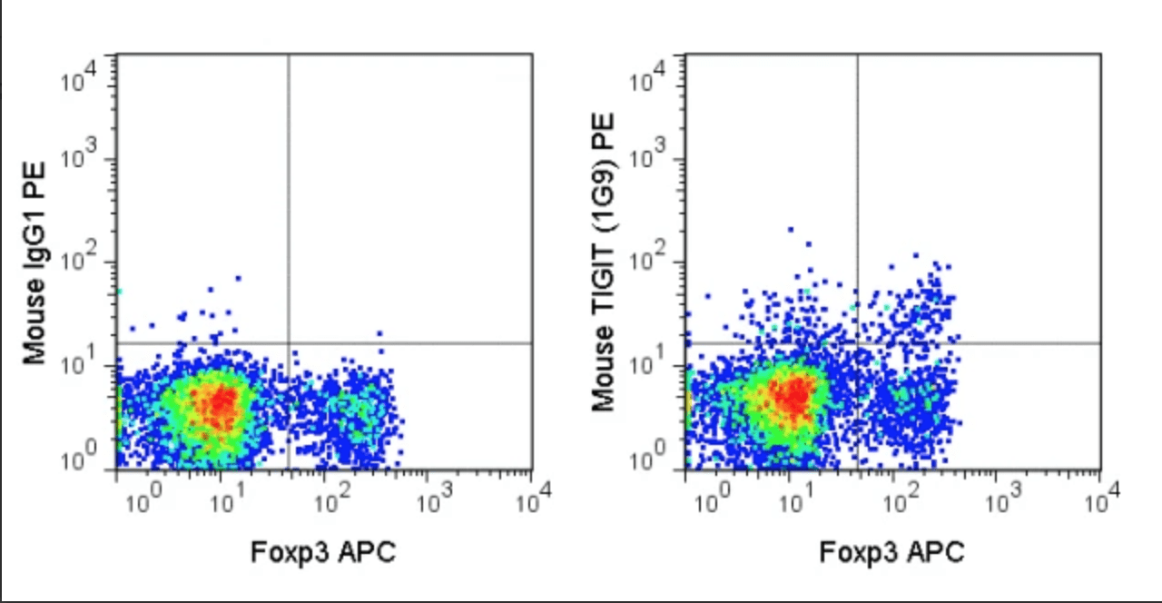
The 1G9 antibody may be used for flow cytometric analysis of TIGIT, which is expressed at very high levels on T regulatory cells (Tregs) and activated conventional T cells, as well as memory T cells and NK cells.
Product Details
| Name | PE Anti-Mouse TIGIT (1G9) |
|---|---|
| Cat. No. | 50-1421 |
| Alternative Names | VSTM3 |
| Gene ID | 100043314 |
| Clone | 1G9 |
| Isotype | Mouse IgG1, kappa |
| Reactivity | Mouse |
| Cross Reactivity | |
| Format | PE |
| Application | Flow Cytometry |
| Citations* | Joller N, Peters A, Anderson AC, and Kuchroo VK. 2012. Immunol. Rev. 248(1):122-139. (Flow cytometry)
Joller N, Hafler JP, Brynedal B, Kassam N, Spoerl S, Levin SD, Sharpe AH, and Kuchroo VK. 2011. J. Immunol. 186: 1338-1342. (Flow cytometry) |
Application Key:FC = Flow Cytometry; FA = Functional Assays; ELISA = Enzyme-Linked Immunosorbent Assay; ICC = Immunocytochemistry; IF = Immunofluorescence Microscopy; IHC = Immunohistochemistry; IHC-F = Immunohistochemistry, Frozen Tissue; IHC-P = Immunohistochemistry, Paraffin-Embedded Tissue; IP = Immunoprecipitation; WB = Western Blot; EM = Electron Microscopy
*Tonbo Biosciences tests all antibodies by flow cytometry. Citations are provided as a resource for additional applications that have not been validated by Tonbo Biosciences. Please choose the appropriate format for each application and consult the Materials and Methods section for additional details about the use of any product in these publications.
[accordions]
[accordion title=”Protocols”]Technical D ate Sheet[/accordion]

