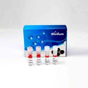
Live-or-Dye NucFix? Red Staining Kit
Live-or-Dye NucFix? Red Staining Kit
Add to cart
Biotium products are distributed only in Singapore and Thailand.
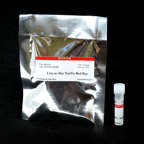
A red fluorescent dye to covalently label dead cells, allowing cells with permeable plasma membranes to be detected in flow cytometry and microscopy.
| SIZE |
CATALOG #
|
PRICE
|
| 50 assays | #32010-T | $64 |
| 200 assays | #32010 | $212 |
PRODUCT ATTRIBUTES
| Apoptosis/viability marker |
Dead cell stain |
|---|---|
| For live or fixed cells |
Covalent & fixable stains, For live/intact cells |
| Assay type/options |
Endpoint assay |
| Detection method/readout |
Fluorescence microscopy, Flow cytometry |
| Fixation options |
Fix after staining (formaldehyde), Fix after staining (methanol), Permeabilize after staining |
| Colors |
Red |
| Excitation/Emission |
520/593 nm (with DNA) |
PRODUCT DESCRIPTION
Live-or-Dye NucFix™ Red is a red fluorescent dye to covalently label dead cells, allowing cells with permeable plasma membranes to be detected in flow cytometry and microscopy.
Features
- Nuclear-specific dead cell stain
- Highly dead-cell specific
- Covalent labeling with reactive dye
- Withstand fixation and permeabilization
- Selective dead-cell staining of mammalian cells, yeast, or gram-negative bacteria
Kit Components
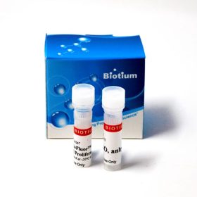
- Live-or-Dye NucFix™ Red Dye
- Anhydrous DMSO
Spectral Properties
- NucFix™ Red Ex/Em 520/593 nm
About Live-or-Dye™
Biotium’s line of Live-or-Dye™ Fixable Viability Staining Kits are designed for discrimination between live and dead cells during flow cytometry or microscopy. Live/dead stains are useful probes to include when analyzing cell surface protein expression by flow cytometry, because they allow intracellular fluorescence signal from dead cells with permeable plasma membranes to be excluded from analysis. In microscopy, live/dead stains allow unambiguous visual discrimination of dead cells.
NucFix™ Red
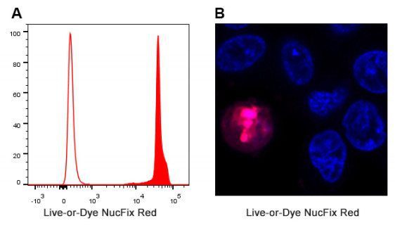
Live-or-Dye NucFix™ Red is a reactive, cell membrane impermeable dye that specifically stains the nuclei of dead cells. The dye is able to enter into dead cells that have compromised membrane integrity and covalently label the cell nucleus, allowing for clear differentiation of live and dead cells by either microscopy or flow cytometry. Unlike other commonly used nuclear stains such as Propidium Iodide (PI) or DRAQ7™, NucFix™ labeling is extremely stable, allowing the cells to be fixed and permeabilized without loss of fluorescence or dye transfer between cells. The Live-or-Dye NucFix™ Red staining protocol has been optimized to maximize live/dead discrimination with minimal live cell staining, in order to prevent interference with immunostaining.
Live-or-Dye NucFix™ Red can also be used as a fixable dead cell stain in gram-negative bacteria. In gram-positive strains there may be live cell staining. Live-or-Dye NucFix™ Red is dead cell specific but not nuclear in yeast. See our Cellular Stains Table for more information on how our dyes stain various organisms.
|
Catalog No. |
Viability Dye |
Laser line |
Emission filter |
Abs/Em Max |
Applications |
|---|---|---|---|---|---|
| 32002, 32002-T | Live-or-Dye™ 350/448 | 355 nm | DAPI or Violet | 347/448 nm | Flow Cytometry |
| 32003, 32003-T | Live-or-Dye™ 405/452 | 405 nm | Pacific Blue® | 408/452 nm | Flow Cytometry |
| 32009, 32009-T | Live-or-Dye™ 405/545 | 405 nm | AmCyan | 395/545 nm | Flow Cytometry |
| 32004, 32004-T | Live-or-Dye™ 488/515 | 488 nm | FITC | 490/515 nm | Flow Cytometry, Microscopy |
| 32012, 32012-T | Live-or-Dye™ 510/550 | 488 nm | Spectral scan | 516/549 nm | Spectral Cytometry |
| 32005, 32005-T | Live-or-Dye™ 568/583 | 488 or 561 nm | PE | 562/583 nm | Flow Cytometry, Microscopy |
| 32006, 32006-T | Live-or-Dye™ 594/614 | 488 or 561 nm | PE-Texas Red® | 593/614 nm | Flow Cytometry, Microscopy |
| 32007, 32007-T | Live-or-Dye™ 640/662 | 633 or 640 nm | APC | 642/662 nm | Flow Cytometry, Microscopy |
| 32013, 32013-T | Live-or-Dye™ 665/685 | 633 or 640 nm | Spectral scan | 667/685 nm | Spectral Cytometry |
| 32008, 32008-T | Live-or-Dye™ 750/777 | 633 or 640 nm | APC-Cy®7 | 755/777 nm | Flow Cytometry |
| 32011, 32011-T | Live-or-Dye™ 787/808 | 785 or 808 nm | APC-Cy®7 or IR800 | 783/803 nm | Flow Cytometry |
| 32010, 32010-T | Live-or-Dye NucFix™ Red | 488 or 532 nm | PE-Texas Red® | 520/593 nm | Flow Cytometry, Microscopy |

 简体中文
简体中文 繁體中文
繁體中文 English
 한국어
한국어 ไทย
ไทย Tiếng Việt