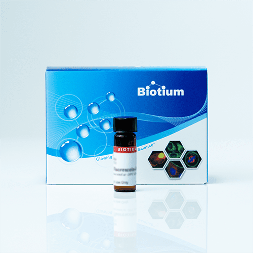
Biotium products are distributed only in Singapore and Thailand.
Visualize Cell Dynamics with DiO: The Classic Lipophilic Tracer
Biotium’s DiO stain offers a reliable and versatile option for labeling cell membranes in your research. This green fluorescent, lipophilic carbocyanine dye is a well-established tool for tracing cellular movement and interactions.
Key Features of DiO:
- Widely Used Tracer: DiO is a popular choice for lipophilic tracing experiments due to its well-documented performance and ease of use.
- Bright Green Fluorescence: The λEx/λEm maxima at 484/501 nm provides clear and distinct labeling for accurate image analysis.
- Compatible with DiI: Combine DiO with DiI for dual-color labeling and colocalization studies, expanding your experimental capabilities.
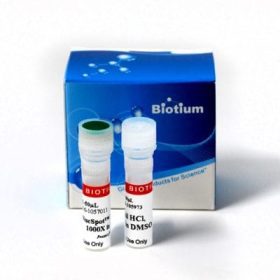
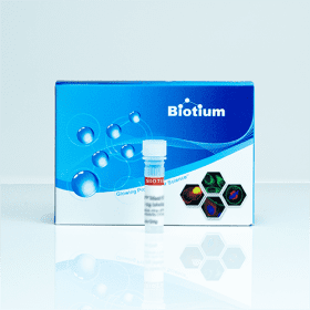
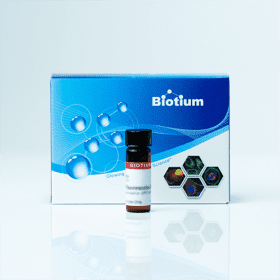
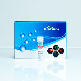
- Multiple Formats: Choose from the versatile DiO powder or the ready-to-use CellBrite™ Green solution for simplified workflow integration.
Please note:
- DiO exhibits lower solubility and a slower lateral diffusion rate compared to newer alternatives like Neuro-DiO.
- For applications requiring superior solubility and faster diffusion, consider exploring Neuro-DiO (Product #60015).
DiO Applications:
- Live cell imaging of cell migration and morphology
- Investigating cell-cell interactions
- Monitoring neurite outgrowth
- Multicolor colocalization studies (in combination with DiI)
Technical Specifications
CAS number: 34215-57-1
Probe cellular localization: Membrane/cell surface, Membrane/vesicular
For live or fixed cells: For fixed cells, For live/intact cells
Assay type/options: Co-cultures, Extended staining (several days to weeks)
Fixation options: Permeabilize before staining, Fix after staining (formaldehyde), Fix before staining (formaldehyde)
Colors: Green
Excitation/Emission: 484/501 nm
λEx/λEm (MeOH): 484/501 nm
Extinction Coefficient (ε): 150,000
Appearance: Yellow orange solid
Solubility: DMF, DMSO, 1:1 DMSO:ethanol (limited)
Storage: 4°C, protected from light
Reference Publications
Alu Konno, Naoya Matsumoto, Yasuko Tomono, and Shigetoshi Okazaki
Pathological application of carbocyanine dye-based multicolour imaging of vasculature and associated structures
Scientific reports vol. 10,1 12613. 28 Jul. 2020
DOI: 10.1038/s41598-020-69394-0
Article Snippet: “We tested several commercially available DiI analogues and successfully introduced three additional dyes with green (DiO), deep red (DiD), and near-infrared (DiR) fluorescence to this technique to increase the number of choices for available colours.”
Elie Beit‐Yannai, Saray Tabak, and W. Daniel Stamer
Physical exosome:exosome interactions
Journal of cellular and molecular medicine vol. 22,3 (2018): 2001-2006.
DOI: 10.1111/jcmm.13479
Article Snippet: “while extracellular vesicles from NTM cells were suspended in the same amount of PBS containing 7.5 μl DiO (3,3′ ‐dioctadecyloxacarbocyanine, perchlorate).”
Janna L. Crossley, et al.
Itaconate-producing neutrophils regulate local and systemic inflammation following trauma
JCI Insight. 2023;8(20):e169208
DOI: https://doi.org/10.1172/jci.insight.169208.
Article Snippet: “Following histopaque-mediated neutrophil isolation from the HO site 7 days after burn/tenotomy in WT mice, cell membranes were labeled with the DiO Lipophilic Carbocyanine fluorescent green dye kit (Biotium, 60011).”
