Biotium products are distributed only in Singapore and Thailand.
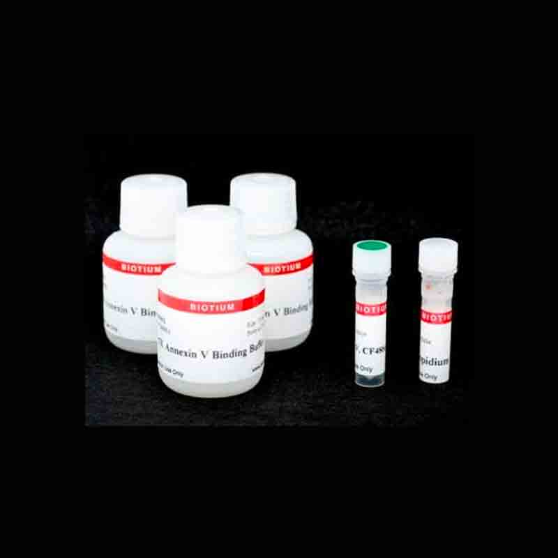
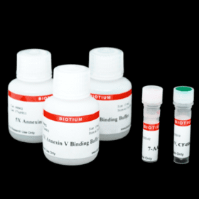
This kit contains CF®488A Annexin V to stain apoptotic cells green and propidium iodide (PI) to stain necrotic cells red for fluorescence microscopy or flow cytometry.
Product Description
This kit contains CF®488A Annexin V for staining apoptotic cells green, and propidium iodide (PI) for staining necrotic cells with red fluorescence, for detection by flow cytometry or fluorescence microscopy.
Features
- Simultaneous detection of apoptotic & necrotic cells
- 15-30 minute staining
- Next-generation CF®488A is brighter and more photostable than FITC
- For fluorescence microscopy or flow cytometry
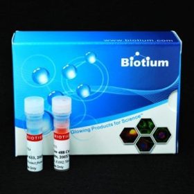
Kit Components
- CF®488A Annexin V (human;recombinant, produced in E. coli)
- Propidium Iodide
- 5X Annexin Binding Buffer
Spectral Properties
- CF®488A Annexin V: Ex/Em 490/515 nm
- PI: Ex/Em 535/617 nm (with DNA)
In apoptotic cells, the phospholipid phosphatidylserine (PS) is translocated from the inner to the outer surface of the plasma membrane, which targets the dying cells for phagocytosis. The human anticoagulant, Annexin V, is a 35 kDa Ca2 -dependent phospholipid binding protein with a high affinity for PS. Annexin V labeled with CF®488A labels apoptotic cells with green fluorescence by binding to PS exposed on the outer leaflet. Our CF®488A dye is superior to fluorescein/FITC because it is not affected by pH and has far better photostability.
Propidium iodide (PI) is impermeant to live cells or early apoptotic cells, but stains necrotic cells and late apoptotic cells with red fluorescence. Both PI and CF®488A Annexin V can be excited using the 488 nm flow cytometry laser line.
See our full selection of Cell Viability & Apoptosis Assays.
Product Attributes
| Apoptosis/viability marker | Phosphatidylserine/Annexin V, Dead cell stain, Apoptosis/necrosis assay |
|---|---|
| For live or fixed cells | For live/intact cells |
| Detection method/readout | Fluorescence microscopy, Flow cytometry |
| Assay type/options | Endpoint assay |
| Colors | Green/Red |
| Product origin | Annexin V (human); recombinant, produced in E. coli |
| Storage Conditions | Store at 2 to 8 °C, Do not freeze, Protect from light |
Reference Publications
Qin, Siyue et al.
Transcriptional factor Nrf2 is essential for aggresome formation during proteasome inhibition
Biomedical reports vol. 11,6 (2019): 241-252.
DOI: https://doi.org/10.3892%2Fbr.2019.1247
Article Snippet: “Cell death-inducing effects of proteasomal inhibition were measured with CF488A-Annexin V (ANXA5) and propidium iodide (PI) Apoptosis kits (cat. no. 30061; Biotium, Inc.) as described previously. A total of 100,000 cells/cm2 were grown in 10 cm dishes. After 24 h, they were treated with DMSO (0.1%) or 2 µM MG132 (in DMSO) for 20 h.”
Kaya-Tilki, Elif, and Miriş Dikmen.
Neuroprotective effects of some epigenetic modifying drugs’ on Chlamydia pneumoniae-induced neuroinflammation: A novel model
PloS one vol. 16,11 e0260633.
DOI: 10.1371/journal.pone.0260633
Article Snippet: “To determine neuronal apoptosis associated with Aβ exposure, 25×103 differentiated SH-SY5Y cells were seeded in 24-well plates and Aβ was applied. After 24 h, cells were trypsinized and staining was performed according to the CF 488A Annexin V and PI Apoptosis Kit (Biotium, USA, Cat. No: 30061) protocol.”
Chenliang Zhang, Ju Gao, Mengen Li, Yongkang Deng and Changan Jiang.
p38δ MAPK regulates aggresome biogenesis by phosphorylating SQSTM1 in response to proteasomal stress
Journal of Cell Science (2018) 131, jcs216671.
DOI: 10.1242/jcs.216671
Article Snippet: “Cell death assay was performed with CF488A-Annexin V (ANXA5) and propidium iodide (PI) apoptosis Kit (Biotium, 30061) as described previously. Briefly, cells grown on 10 cm dishes were treated with DMSO or 2 µM MG132 for 18 h, then harvested by digestion with 0.05% trypsin-EDTA solution.”

