1.Microcarrier culture
Prepare the digested cells, add the previously prepared microcarriers to a square flask or reactor (microcarrier addition 3-5g/L), start culturing under certain conditions, and observe the attachment of cells after 4 hours. If the cells adhere well, continue to follow the culture and observe the cell growth.
Key points of microcarrier culture operation
(1) Early stage of culture: Ensure that the medium and microspheres are at a stable pH and temperature level, and inoculate cells (logarithmic growth phase, not stable phase) to 1/3 of the final volume of the culture medium to increase cells and microcarriers Opportunity for contact. The microcarrier content of 2-3g/L is often used, and higher microcarrier concentrations require environmental control or frequent liquid replacement.
Since animal cells have no cell wall and are sensitive to shearing force, it is impossible to increase the contact probabilities by increasing the stirring speed. The usual method of operation is to use a low stirring speed during the adherence phase and stir at all times. The stirring process is stopped a few hours later as the cells are become attached to the surface of the microcarrier. Instead a low speed is set as the cells enter the culture phase. The stirring of microcarrier culture is very slow, the maximum speed is 75r/min.
(2) After the adherence stage (3-8h), slowly add the culture medium to the working volume, and increase the stirring speed to ensure complete homogeneous mixing.
(3) Culture maintenance period: cell count (nucleus count), glucose measurement and cell morphology microscopy. As the cells proliferate, the microspheres become heavier and heavier, and the stirring rate needs to be increased(Photos of carrier cells at different culture times were shown in Figure 1 to Figure 3)After about 3 days, the culture medium begins to be acidic and needs to be changed. Specific method: stop stirring, let the Cell Dex settle for 5 minutes, discard the appropriate volume of culture solution, slowly add fresh culture solution (37°C), and restart stirring.
(4) Harvesting cells (This digestion method may require the use of off-line sterilization of pancreatin digester)
A. The cells to be digested are fed into the nearly sterilized microcarrier digester through a sterile pipeline. The culture medium is discharged from the digester through the filter system of the microcarrier digester, and the microcarrier with overgrown cells is retained above the microcarrier digester.
B. Add 1~5 times the volume of calcium-magnesium free PBS of the remaining carrier into the microcarrier digester, open the stirring system of the digester, make PBS fully contact with the microcarrier, and discharge calcium-magnesium free PBS after continuous stirring for 3~5min.
C. Repeat cleaning with PBS twice according to the same method as B, and discharge calcium-magnesium free PBS
D. PBS solution containing 0.02%EDTA was added to the microcarrier cleaned by PBS, and stirred continuously at 37℃ for about 10 minutes before discharging
E. Add trypsin containing 0.125% trypsin and 0.02%EDTA 75-125r/min and stir quickly for 20-30min (digestion time is observed under a microscope and digestion stops when there are no cells on the carrier).
F. After digestion, the digested cells are transferred to a larger bioreactor through the drain tube of the digester.
(5) Scale-up of microcarrier culture: It can be scaled up by increasing the content of microcarriers or the culture volume. The use of aneuploidy or primary cell culture to produce vaccines and interferon has been amplified to more than 4000L.
2.Storage
Store in a dry, ventilated and clean place at 4~25°C.
3. Transportation
Avoid sunlight, rain, and heavy pressure during transportation, and it is strictly forbidden to transport it with toxic and hazardous materials.
4. Caution
Avoid to contact oxidants.
5.Order instruction
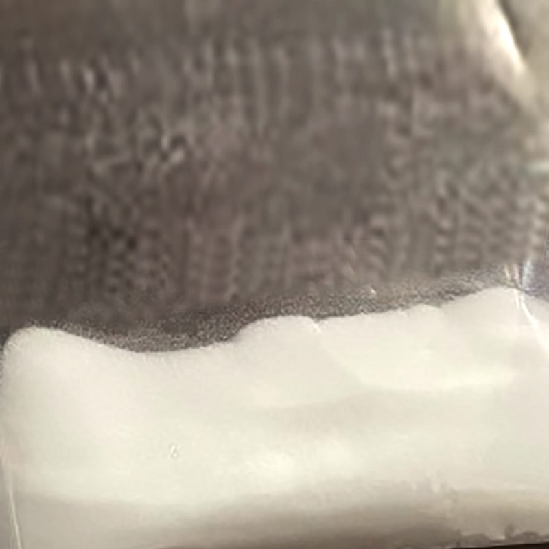
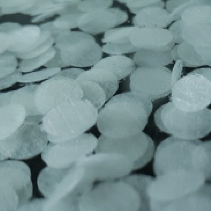
Celldex is supplied as a dry powder and must be hydrated and sterilized before use.
| Product name | Series No. | Packing |
| Celldex | SABMCD02.0250 | 250g |
| SABMCD02.0500 | 500g | |
| SABMCD02.1000 | 1kg | |
| SABMCD02.5000 | 5kg | |
| SABMCD02.10000 | 10kg |
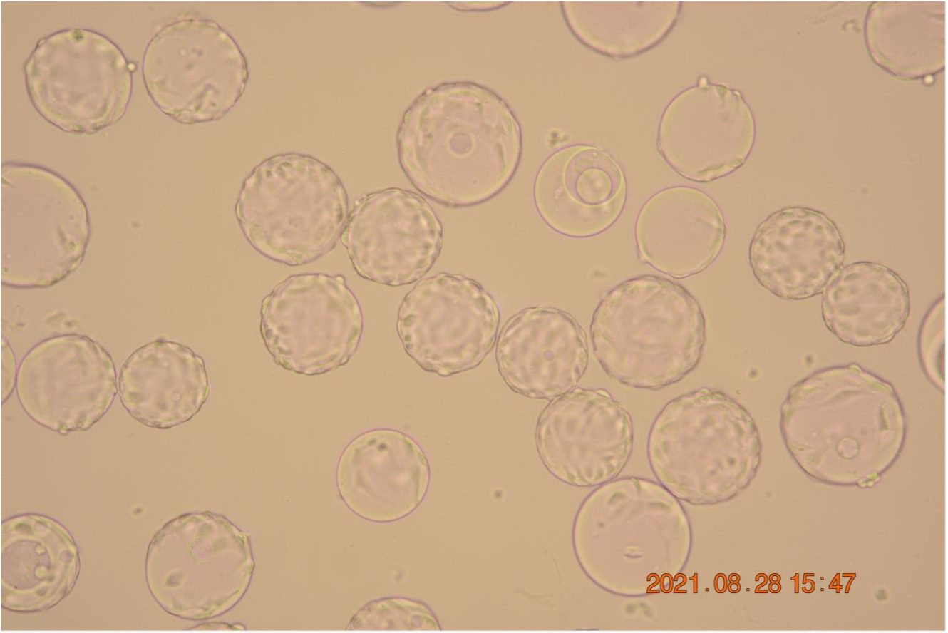
Pic 1. Photomicrograph of Vero cells cultured in CellDex1 for 24h
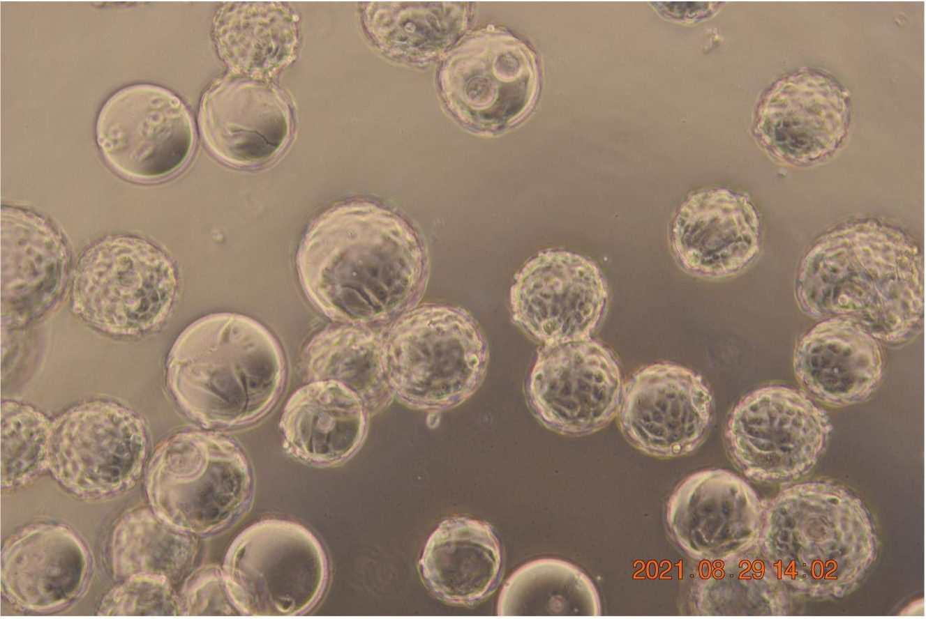
Pic 2. Photomicrograph of Vero cells cultured in CellDex1 for 48h(320 x)
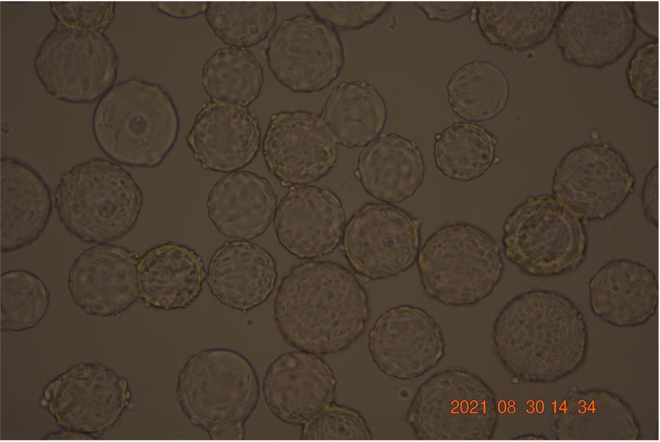
Pic3. Photomicrograph of Vero cells cultured in CellDex1 for 72h(320 x)

