Biotium products are distributed only in Singapore and Thailand.
A convenient assay for quantifying apoptotic (green) and necrotic (red) cells within the same cell population by flow cytometry or fluorescence microscopy.
Product Description
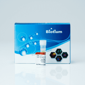
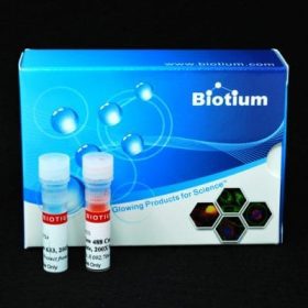
The Apoptosis and Necrosis Quantitation Kit Plus provides a convenient assay for quantifying apoptotic (green) and necrotic (red) cells within the same cell population by flow cytometry or fluorescence microscopy.
Features
- Simultaneous detection of apoptotic & necrotic cells
- 15-30 minute staining
- Next-generation CF®488A is brighter and more photostable than FITC
- EthD-III is a superior alternative to PI and EthD-I
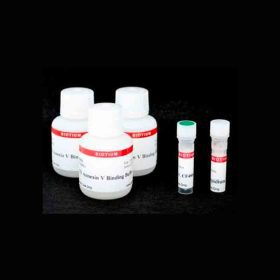
Kit Components
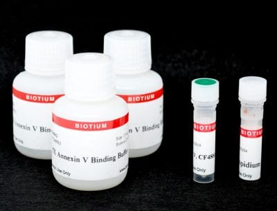
- CF®488A Annexin V (human; recombinant, produced in E. coli)
- Ethidium Homodimer III (EthD-III)
- 5X Annexin Binding Buffer
Spectral Properties
- CF®488A Annexin V: Ex/Em 490/515 nm
- EthD-III: Ex/Em 279, 522/593 nm (with DNA)
Apoptosis and necrosis are two major processes by which cells die. Apoptosis is an active, genetically regulated disassembly of the cell from within. During apoptosis, phosphatidylserine (PS) is translocated from the inner to the outer leaflet of the plasma membrane for phagocytic cell recognition. The human anticoagulant Annexin V is a 35 kDa, Ca2+-dependent phospholipid-binding protein with high affinity for PS. Annexin V labeled with CF®488A stains apoptotic cells green by binding to PS exposed on the cell surface. CF®488A is spectrally similar to fluorescein (FITC), with much brighter and more photostable fluorescence.
Necrosis normally results from severe cellular insult. Both internal organelle and plasma membrane integrity are lost, resulting in the release of cellular contents into the surrounding environment. Ethidium Homodimer III (EthD-III) is a highly positively charged nucleic acid probe, which is impermeant to live cells, but stains cells with compromised membrane integrity with red fluorescence. EthD-III is a superior alternative to Propidium Iodide (PI) or Ethidium Homodimer I because of its significantly higher affinity for DNA and higher fluorescence quantum yield. The Apoptosis & Necrosis Quantitation Kit Plus provides a convenient assay for quantifying apoptotic (green) and necrotic (red) cells within the same cell population by flow cytometry or fluorescence microscopy.
See our full selection of Cell Viability & Apoptosis Assays and Viability & Apoptosis Assays for Flow Cytometry.
Product Attributes
| Apoptosis/viability marker | Phosphatidylserine/Annexin V, Dead cell stain, Apoptosis/necrosis assay |
|---|---|
| For live or fixed cells | For live/intact cells |
| Detection method/readout | Fluorescence microscopy, Flow cytometry |
| Assay type/options | Endpoint assay |
| Colors | Green/Red |
| Product origin | Annexin V (human); recombinant, produced in E. coli |
| Storage Conditions | Store at 2 to 8 °C, Do not freeze, Protect from light |
Reference Puclications
Czuczi, Tamás et al.
Synthesis and Antiproliferative Activity of Novel Imipridone-Ferrocene Hybrids with Triazole and Alkyne Linkers
Pharmaceuticals (Basel, Switzerland) vol. 15,4 468.
DOI: 10.3390/ph15040468
Article Snippet: “PANC-1 and A2058 cells were seeded in eight-well Ibidi® μ-Slide microscopic slides (5 × 10³ cells/well) and were allowed to adhere for 48 h. Apoptosis and Necrosis Quantitation Kit Plus (Biotium, cat. no.: 30065) was used, according to the manufacturer’s instructions.”
Yang, Jiang et al.
Gold/alpha-lactalbumin nanoprobes for the imaging and treatment of breast cancer
Nature biomedical engineering vol. 4,7 (2020): 686-703.
DOI: 10.1038/s41551-020-0584-z
Article Snippet: “Apoptosis and necrosis were estimated using the Apoptosis and Necrosis Quantification Kit plus (30065, Biotium) according to the manufacturer’s instructions. In brief, trypsinized MDA-MB-231 cells after treatment were resuspended in binding buffer at 3 × 10⁶ cells per ml.”
Murányi J, Duró C, Gurbi B, Móra I, Varga A, Németh K, Simon J, Csala M, Csámpai A.
Novel Erlotinib–Chalcone Hybrids Diminish Resistance in Head and Neck Cancer by Inducing Multiple Cell Death Mechanisms
International Journal of Molecular Sciences. 2023; 24(4):3456.
DOI: https://doi.org/10.3390/ijms24043456
Article Snippet: “Apoptosis and Necrosis Quantitation Kit Plus (Biotium, cat. no.: 30065) was used according to the manufacturer’s instructions. For nuclear staining of live cells, DRAQ5 was used (5 µM, 30 min). Images of cells were acquired with a confocal laser microscope”

