APO-DIRECT™ Kit
$440.00
Add to cart
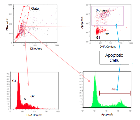
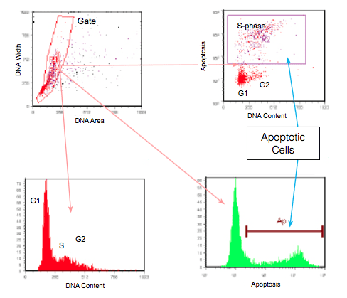
The fragmentation of genomic DNA by cellular nucleases during the later stages of apoptosis is also one of the most easily measured features of apoptotic cells. Nuclease activity generates DNA fragments ranging from ~300 bp to 50 bp in length, resulting in a typical DNA ‘laddering’ appearance when analyzed by agarose gel electrophoresis. These fragments have exposed 3’-hydroxyl (OH) ends which can be labeled with deoxyuridine triphosphates (dUTP). An enzyme, terminal deoxynucleotidyl transferase (TdT), is used to catalyze the template-independent addition of fluorescent tagged dUTP to the 3’-OH ends of double or single stranded DNA. This method is often called TUNEL (terminal deoxynucleotidyl transferase dUTP nick end labeling) or end labeling.
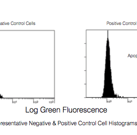
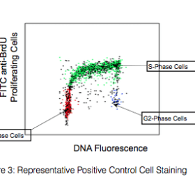
With the APO-DIRECT™ Kit, cells are stained with FITC-labeled dUTP in a single step. Samples can then be analyzed via flow cytometry. Samples that are apoptotic will stain brightly due to the substantial number of exposed 3’-OH sites, while cells that are non-apoptotic will not have incorporated significant amounts of FITC-dUTP and will stain dimly.
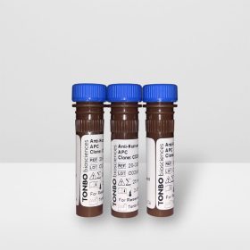
The APO-DIRECT Kit is shipped in one container and consists of two packages plus instruction manual. Upon arrival one should be stored at 2-8°C and the other at -20°C.
Product Details
| Name | APO-DIRECT™ Kit |
|---|---|
| Cat. No. | TNB-6611 |
| Clone | N/A |
| Isotype | N/A |
| Format | FITC |
| Application | Flow Cytometry |
| Citations* | Li X, Traganos F, Melamed MR and Darzynkiewicz Z. 1995. Cytometry. 20(2): 172-180.
Tomei LD and Cope FD, eds. 1991. Current Communications in Cell and Molecular Biology. 3: 47-60. Cold Springs Harbor, NY. Arends MJ, Morris RG and Wyllie AH. 1990. Am J Pathol. 136(3): 593-608. Darzynkiewicz Z, Crissman HA and Robinson JR, eds. 1994. Methods in Cell Biology: Flow Cytometry Second Edition. 41: 15-38. Academic Press Inc. Eschenfeldt WH, Puskas RS and Berger SL. 1987. Methods Enzymol. 152: 337-342. Darzynkiewicz Z, Bruno S, Del Bino G, Gorczyca W, Hotz MA, Lassota P and Traganos F. 1992. Cytometry. 13(8): 795-808. |
Application Key:FC = Flow Cytometry; FA = Functional Assays; ELISA = Enzyme-Linked Immunosorbent Assay; ICC = Immunocytochemistry; IF = Immunofluorescence Microscopy; IHC = Immunohistochemistry; IHC-F = Immunohistochemistry, Frozen Tissue; IHC-P = Immunohistochemistry, Paraffin-Embedded Tissue; IP = Immunoprecipitation; WB = Western Blot; EM = Electron Microscopy
*Tonbo Biosciences tests all antibodies by flow cytometry. Citations are provided as a resource for additional applications that have not been validated by Tonbo Biosciences. Please choose the appropriate format for each application and consult the Materials and Methods section for additional details about the use of any product in these publications.


 简体中文
简体中文 繁體中文
繁體中文 English
 한국어
한국어 ไทย
ไทย Tiếng Việt