- Your cart is empty
- Continue Shopping
Tips for Staining 3D Organoids and Spheroids

Tips for Staining 3D Organoids and Spheroids
- Atlantis Bioscience
- Blog
- Reading Time: 7 minutes
Organoids and spheroids represent a breakthrough in biological research, offering advanced 3D models that more accurately mimic the architecture and function of human tissues than traditional 2D cultures. These three-dimensional structures are created by cultivating cells in specialised environments that promote self-organisation, forming complex tissues with characteristics resembling in vivo organs.
What are the differences between Organoids and Spheroids?
- Organoids are miniaturised versions of organs, grown from stem cells or organ-specific progenitors, and are capable of developing multiple cell types and even some functional properties of their respective organs. Organoids derived from tissues like the liver, kidney, brain, or intestines provide researchers with a powerful model to study organ development, disease progression, and even responses to drugs.
- Spheroids, on the other hand, are more simplified 3D cell aggregates that lack the tissue complexity seen in organoids but still provide an essential bridge between 2D cell cultures and more complex in vivo models. Spheroids can be formed from cancer cells, allowing researchers to study tumour behaviour, drug resistance, and the interaction between cancer cells and their microenvironment.
| Feature | Organoids | Spheroids |
| Definition | Mini-organs with structural/functional resemblance to real organs. | Simplified 3D cell aggregates. |
| Origin | Derived from stem cells or progenitors. | Formed from various cell types (e.g., tumour cells). |
| Complexity | High, with multiple cell types and layers. | Low, homogeneous cell clusters. |
| Structure | Resembles organ tissue with distinct regions. | Uniform, less organised structure. |
| Size | Larger, varies by organ type. | Smaller, generally a few hundred microns. |
| Functionality | Mimics some organ functions (e.g., secretion, signalling). | Lacks organ-like functionality. |
| Culture Requirements | Requires complex ECM scaffolds (e.g., Matrigel). | Easier, typically grown in suspension. |
| Applications | Disease modelling, drug screening, regenerative medicine. | Tumour biology, drug testing, cancer research. |
| Examples | Brain, liver, and intestinal organoids. | Cancer and hepatocyte spheroids. |
Both organoids and spheroids are transforming areas of research such as cancer biology, drug discovery, toxicology, and regenerative medicine. By offering a more physiologically relevant model system, they enable researchers to:
- Study complex cell-to-cell and cell-to-matrix interactions.
- Model diseases more accurately, including patient-specific conditions.
- Test drug efficacy and toxicity in a more predictive manner before moving to animal models or clinical trials.
Their growing importance in research highlights the need for precise methods to visualise and study these 3D structures, making staining and imaging critical to gaining meaningful insights into their biology.

Staining 3D Organoids and Spheroids
Staining 3D organoids and spheroids for research is a complex process due to their size, dense cellular composition, and the need to preserve their structural integrity. This section highlights the main challenges and offers practical tips to optimise staining and imaging.
1. Challenges in Staining 3D Structures
- Limited Penetration of Stains: Due to the size and density of organoids and spheroids, stains may not penetrate uniformly, particularly in the core regions. To ensure even distribution, optimize the concentration of stains and antibodies and extend incubation times (up to 72 hours if needed). Gentle agitation can also help improve stain penetration.
- High Background Signal: Autofluorescence from the extracellular matrix (ECM) or cell debris can interfere with specific staining, resulting in high background noise. Optical clearing methods or autofluorescence quenching agents can help reduce this issue, improving signal-to-noise ratios.
- Preserving 3D Structure: Organoids and spheroids are delicate, and staining procedures can risk damaging or deforming their structures. Gentle handling with soft pipetting, embedding in hydrogels, and low-speed centrifugation are effective methods for maintaining the integrity of these 3D cultures during staining.
- Fixation Issues: Overfixation can block stain penetration, while underfixation can result in weak signals. Fixation protocols should be optimised, typically using 4% paraformaldehyde for 15-60 minutes, depending on the size of the organoid or spheroid. Adjust the protocol based on preliminary testing.
2. Tips for Effective Staining
- Optimise Permeabilisation: Effective permeabilisation is essential for allowing stains to penetrate deeper into organoids. Using agents like Triton X-100 (0.1-0.5%) with optimised concentrations and incubation times can enhance staining without damaging the structures.
- Prolong Incubation Times: Given the size of 3D models, longer incubation times are often necessary. Prolonging stain incubation for 24-72 hours can ensure the dye or antibody penetrates into the inner layers of the spheroid or organoid.
- Multi-Step Gentle Washing: Gentle, multi-step washing is essential to remove excess staining reagents without disturbing the delicate 3D structures. Use low-speed centrifugation or gravity settling during washing steps to prevent loss or damage.
3. Adapting Imaging Techniques
- Confocal vs. Light-Sheet Microscopy: Traditional confocal microscopy works well for sectioning through thin slices, but it can be limited for thicker 3D structures like organoids. Light-sheet or two-photon microscopy offers better visualisation for thicker samples and is more effective for capturing the entire structure in 3D.
- Z-Stacking and 3D Reconstruction: For a complete visualisation of organoids and spheroids, Z-stacking is essential. By acquiring images in multiple focal planes, researchers can reconstruct the 3D structure. Post-acquisition image reconstruction and analysis software (e.g., Imaris, Fiji) can aid in this process.
- Optical Clearing: Organoids are often opaque, which can reduce imaging clarity. Optical clearing techniques enhance transparency, allowing for better visualisation of internal structures without affecting staining.
4. Common Pitfalls and How to Avoid Them
- Overstaining: Excessive staining can obscure specific signals and lead to uneven labelling, particularly in the outer layers of spheroids. Titrate stains carefully and run controls to determine optimal concentrations.
- Inadequate Mounting Media: The refractive index mismatch between the mounting medium and the stained sample can lead to poor image quality. Using mounting media designed for 3D tissues will ensure optimal imaging and minimal distortion.
Basic Protocol for Staining Spheroids
Proper staining techniques are crucial for visualising cellular structures and assessing the expression of specific proteins within spheroids. This section outlines a brief protocol for staining spheroids, encompassing fixation, staining, and imaging.
Steps:
1. Fixation
- Gently collect spheroids using a wide-bore pipette tip and transfer to a 1.5 mL centrifuge tube. Allow spheroids to sediment and aspirate supernatant carefully.
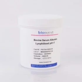
- Tip: Pre-coating the tips and centrifuge tube with 1% BSA in PBS may help ensure maximal transfer efficiency.
- Fix spheroids in 4% paraformaldehyde (PFA) for 15-20 minutes at room temperature, and quench with 50 mM Tris/HCl (pH 7.4) for 30 minutes. Depending on the size of the spheroid, larger spheroids may require longer fixation time.
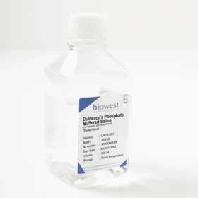
- After fixation, wash spheroids 3 times with PBS (5 minutes each wash).
- Note: The cells can be maintained at 4 °C for long-term storage (>1 year).
2. Permeabilisation
- Incubate spheroids in 0.1-1.0% Triton X-100 (or an alternative permeabilisation agent) in PBS for 3 hours at room temperature. Adjust permeabilisation time based on spheroid density and the size of the molecule you want to stain.
- Wash spheroids again with PBS (3 washes, 5 minutes each).
3. Blocking
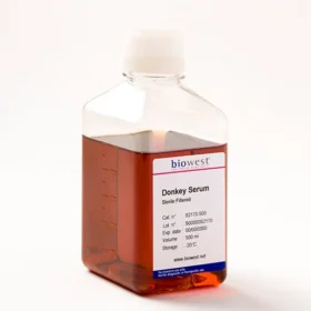
- Incubate spheroids in blocking buffer (e.g., 1% BSA, and 1% donkey serum in PBS) for 1 hour at room temperature to reduce non-specific staining.
- Tip: Use animal serum in which the secondary antibodies were raised to reduce background.
- Note: BSA usually works well for the blocking step, but in case of high background noise, perform an empirical test to obtain the best possible results for a given combination of antibodies.
- Gently agitate or rock during the incubation to ensure even distribution of the blocking buffer.
4. Staining
- Prepare your staining solution (e.g., primary antibody, nuclear dye, or fluorescent probe) diluted in PBS with blocking buffer.
- Incubate spheroids in the staining solution for 24-72 hours at 4°C. Prolonged incubation ensures that the stain reaches the innermost cells of the spheroid.
- Optionally, keep spheroids under gentle agitation (e.g., on a rocker or rotating platform) to enhance penetration.
5. Washing
- Wash the spheroids 3-5 times in PBS to remove unbound stain. Ensure that the washes are gentle to preserve spheroid integrity.
6. Conterstaining (if needed)
- If a secondary antibody or counterstain is needed (e.g., for detecting a primary antibody or staining additional markers), incubate spheroids with the secondary staining solution for an additional 24-48 hours at 4°C.
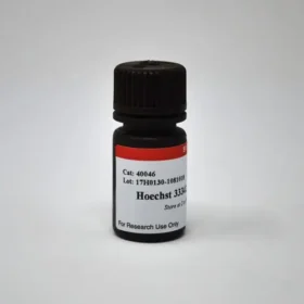
- Add Hoechst 33342 and incubate for another 2 hours at 4 °C
- Wash again 3-5 times with PBS.
- Note: The protocol can be paused here, and the samples can be stored at 4 °C for several months, protected from light.
7. Mounting
- Transfer stained spheroids to a glass slide or imaging chamber with mounting media designed for 3D imaging. Ensure the medium is compatible with your imaging method and minimises autofluorescence.
8. Microscopy
- Image the stained spheroids using confocal, light-sheet, or two-photon microscopy for optimal visualisation. Ensure that the acquisition settings (Z-stacking, laser power) are adjusted for 3D samples.
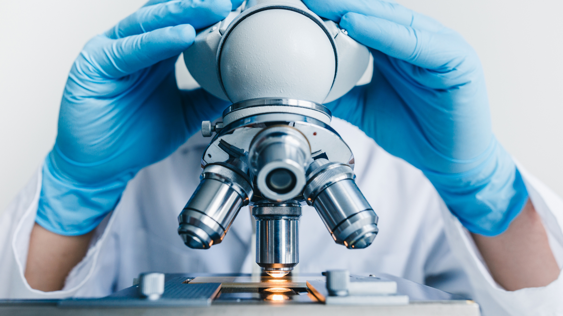
Stains for Spheroids, 3D Cultures, or Matrigel® Cultures
Unlike 2D cultures, spheroids present challenges in terms of depth and organisation, making it difficult to assess cell viability, proliferation, or specific biological markers without using dyes or stains. By employing appropriate stains, researchers can differentiate between live and dead cells, track cellular components like membranes and mitochondria, and even monitor processes like apoptosis. This enhances our ability to understand cell behaviour, organisation, and responses to treatments within the 3D environment.
| Stains/Dyes | Target | Fixable? | Applications/Notes |
| Hoechst Dyes | All cell nuclei | Yes | Commonly used to stain cells in Matrigel® or spheroids |
| NucSpot® Live Cell Nuclear Stains | All cell nuclei | Yes | Long-term live cell imaging or fixed cell staining |
| CellBrite® Cytoplasmic Membrane Dyes | Cell membranes | Yes | Lipophilic carbocyanine dye have been used for labeling cells before or after spheroid formation |
| MitoView™ Dyes | Mitochondria | No | Long-term live cell imaging |
| NucView® 488 Caspase-3 Substrate | Apoptotic Cells | Yes | Long-term live cell imaging |
| Calcein-AM | Viable cells (whole cell interior) | No | Commonly used to stain cells in Matrigel® or spheroids |
| ViaFluor® SE Cell Proliferation Kits | Whole cell interior | Yes | Can be used to label cells before seeding in Matrigel® or spheroids |
| LipidSpot™ Lipid Droplet Stains | Lipid droplets | Yes | Rapidly and specifically stain lipid droplets |
References:
Bergdorf KN, Phifer CJ, Bechard ME, et al. Immunofluorescent staining of cancer spheroids and fine-needle aspiration-derived organoids. STAR Protoc. 2021;2(2):100578. Published 2021 May 31. doi:10.1016/j.xpro.2021.100578
Genenger B, McAlary L, Perry JR, Ashford B, Ranson M. Protocol for the generation and automated confocal imaging of whole multi-cellular tumor spheroids. STAR Protoc. Published online June 9, 2023. doi:10.1016/j.xpro.2023.102331
Gonzalez AL, Luciana L, Le Nevé C, et al. Staining and High-Resolution Imaging of Three-Dimensional Organoid and Spheroid Models. J Vis Exp. 2021;(169):10.3791/62280. Published 2021 Mar 27. doi:10.3791/62280
Haspels B, Kuijten MMP. Protocol for formation, staining, and imaging of 3D breast cancer models using MicroTissues mold systems. STAR Protoc. Published online September 20, 2024. doi:10.1016/j.xpro.2024.103250
Smyrek I, Stelzer EH. Quantitative three-dimensional evaluation of immunofluorescence staining for large whole mount spheroids with light sheet microscopy. Biomed Opt Express. 2017 Jan 3;8(2):484-499. doi: 10.1364/BOE.8.000484.
CONTACT

QUESTIONS IN YOUR MIND?
Connect With Our Technical Specialist.

KNOW WHAT YOU WANT?
Request For A Quotation.
OTHER BLOGS YOU MIGHT LIKE
HOW CAN WE HELP YOU? Our specialists are to help you find the best product for your application. We will be happy to help you find the right product for the job.

TALK TO A SPECIALIST
Contact our Customer Care, Sales & Scientific Assistance

EMAIL US
Consult and asked questions about our products & services

DOCUMENTATION
Documentation of Technical & Safety Data Sheet, Guides and more...

 简体中文
简体中文 繁體中文
繁體中文 English
 한국어
한국어 ไทย
ไทย Tiếng Việt