- Your cart is empty
- Continue Shopping

Imagine dedicating years of research to investigating a disease or testing a novel therapeutic, only to discover that your results were based on misidentified or contaminated cell lines. This issue, affecting up to 36% of cell lines used in scientific studies, threatens research validity and wastes valuable resources. Given the critical role cell lines play in biomedical research, thorough authentication and validation are essential to ensure reproducibility and scientific integrity.
This blog will explore the essential steps for validating a new cell line to ensure that your research is built on a solid foundation, free from the pitfalls of contamination and misidentification.
Regular testing of cell lines is crucial for maintaining the integrity of your research. Testing helps prevent contamination, misidentification, and genetic drift—common issues that can compromise experimental results. Below are the key moments when cell line testing should be prioritised:
Whenever you receive a new cell line, whether from another lab or a commercial supplier, it is essential to confirm its identity before any experimental work begins. Similarly, testing cell lines before banking them for future use ensures that you are preserving a verified, uncontaminated line from the start.
Regularly scheduled testing is essential for cell lines in continuous use. Testing at defined intervals, such as after a set number of passages, helps detect genetic changes, contamination, or drift that may affect cell behaviour. Establishing a routine QC schedule ensures stability throughout your experiments, helping to catch issues early before they impact results.
Many high-impact journals now require proof of cell line authentication as part of their submission guidelines. For example, Nature Publishing Group and American Association for Cancer Research mandate documentation of the authentication of all cell lines used in their study. Ensuring your cell lines are validated before submission helps protect the credibility of your research and aligns with these journal requirements.
If your cells suddenly exhibit abnormal behaviour—such as accelerated growth, altered morphology, or unexpected responses to treatments—this could be a sign of contamination or misidentification. Testing at the first sign of unusual results can help identify whether contamination or genetic drift is affecting your culture.
Introducing stable transfection, gene editing, or drug selection can lead to the emergence of variant sublines or more robust cell types that outcompete the intended population. Testing after these modifications ensures the continued authenticity and stability of your cell line.
If you are working with a novel or unique cell line, it is critical to establish a genetic profile as early as possible. This baseline data allows you to monitor the cell line over time for any changes or contamination, ensuring the genetic integrity of your cell model remains intact for future experiments.
When validating a new cell line for research use, adhering to proper safety, procedural, and operational protocols is critical. Below are the essential steps to ensure the integrity and safety of your research:
Understand and comply with the safety regulations of your institution. Consult your Biological Safety Officer or advisor to ensure you are following guidelines on handling hazardous materials, waste disposal, and maintaining a sterile work environment.
Always treat human tissue and associated cell cultures as potentially infectious. Wear appropriate personal protective equipment (PPE) and work in biosafety cabinets to minimise the risk of exposure to harmful pathogens.
Before starting any procedure, confirm disposal routes for all types of laboratory waste, including biological materials, chemicals, and sharps. Ensure that your lab complies with local regulations, such as autoclaving biological waste or segregating hazardous chemicals, to maintain lab and environmental safety.
All lab personnel should be properly trained in standard operating procedures (SOPs), biosafety protocols, and emergency responses. Regular refresher training should be documented, ensuring competency and reducing errors that could compromise safety or experimental outcomes.
Always source media, serum, and other reagents from validated, reliable suppliers. Consistency in the quality of reagents is essential to reduce variability and contamination in cell cultures, ensuring reproducible results.
Perform media preparation in a dedicated, sterile area, separate from cell culture handling. This reduces the risk of cross-contamination between the reagents and the living cell cultures, preserving the integrity of your experiments.
Keep detailed records of batch numbers for media, reagents, and sera used in your cell cultures. This traceability allows you to identify and address any inconsistencies or contamination issues that arise during your experiments.
SOPs should be in place for all routine laboratory tasks, such as cell culture, media preparation, cryopreservation, and equipment maintenance. Adherence to SOPs ensures consistency, reproducibility, and adherence to best practices throughout your research.
Ensure all laboratory equipment, including biosafety cabinets, incubators, autoclaves, and water filtration systems, is regularly serviced and calibrated. Well-maintained equipment is crucial for maintaining a controlled environment and preventing contamination or experimental failure.
Before using a new cell line, inspect it under an inverted phase-contrast microscope. For routine culture, inspect cells daily and refer to reference images to track growth patterns and morphology. Understanding your cell line’s normal behaviour will help you quickly identify contamination or other issues.
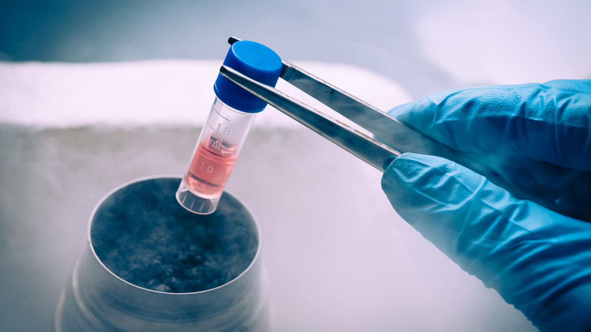
Freeze new cell lines as early as possible, preferably at a low passage number, after clearing quarantine. This ensures that genetically stable cells are preserved for future use. If freezing must occur before clearing quarantine, handle these cell lines as if they are contaminated and follow strict containment protocols.
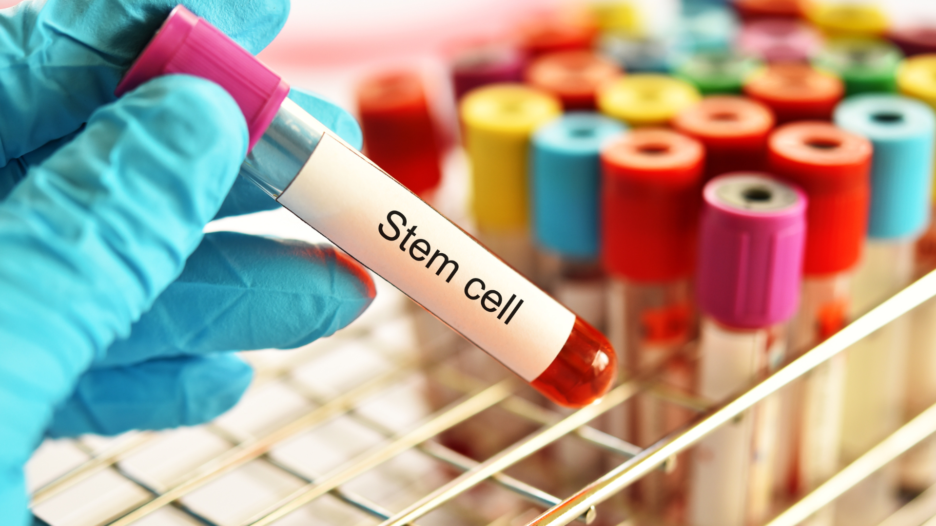
To ensure the reliability and integrity of research, it’s essential to perform routine cell line authentication tests. Key methods include Short Tandem Repeat (STR) profiling for human cell lines, Single Nucleotide Polymorphism (SNP) analysis for genetic stability, and karyotyping or Fluorescence In Situ Hybridisation (FISH) to detect chromosomal abnormalities. Regular mycoplasma testing is also crucial, as contamination can alter cell behaviour. Researchers should authenticate cell lines when acquiring new cultures, before freezing stocks, and periodically throughout experiments to ensure consistent performance and prevent cross-contamination. By incorporating these tests into routine quality control, you safeguard the validity of experimental results.
Cellular morphology refers to the observation of cells under a microscope, providing a quick and direct way to assess the health and status of a culture. The appearance of cells can change based on several factors, including cell type, plating density, medium composition, and the health or differentiation state of the cells. Morphology can vary between different cell lines and within the same cell line under different conditions.
To obtain reliable information, it’s important to regularly observe cultures at both high and low densities. Frequent brief observations allow researchers to monitor any sudden or gradual changes in cell appearance. If the morphology differs from the expected pattern, it can indicate potential problems, such as contamination or poor health of the cells. Researchers should capture and maintain cell morphology images over time to have reference points for comparison. If unusual morphological features appear, it’s crucial to investigate further.
Analysing cell growth through growth curves is another essential method for monitoring the health and consistency of cell cultures. Growth curve analysis involves measuring the proliferation rate of cells over time. A healthy cell line typically exhibits predictable growth patterns, and any sudden changes in growth rate—either an unexpected increase or decrease—can signal an underlying issue with the culture, such as contamination, genetic drift, or poor medium quality.
By establishing a growth baseline, researchers can determine key parameters like population doubling times, optimal subculturing schedules, and plating efficiencies. Monitoring cell growth also helps identify the best points in the cell cycle for harvesting cells for experiments. Consistent growth behaviour is a strong indicator of a stable and healthy cell line, and growth curves should be regularly updated when preparing for critical assays or when changes are made to the culture conditions.
Isoenzyme analysis is an effective method for verifying the species of origin of a cell line. Different species produce distinct isoenzymes, which can be separated based on their electrophoretic properties. By comparing the distribution patterns of specific enzyme groups, isoenzyme analysis can confirm the species identity of the cell line and detect contamination by another species.
Cytogenetic analysis is vital for verifying the chromosomal integrity and genetic stability of cell lines. Two key methods used are karyotyping and FISH.
STR profiling, also known as DNA fingerprinting, is a powerful tool used to verify the identity of human cell lines. STR profiling works by amplifying specific regions, typically 2–6 base pairs in length, known as STR loci, which vary greatly between individuals. Each cell line has a unique STR profile, allowing researchers to establish a genetic “fingerprint” that can be stored and used for future comparisons.
This technique helps prevent the use of misidentified or contaminated human cell lines, ensuring the integrity of research. STR profiling is often performed using multiplex PCR to amplify multiple polymorphic markers, which are then analysed using software to generate a unique DNA profile for each cell line. The profile acts as a baseline for ongoing quality control checks, ensuring the line remains consistent and unaltered over time.
SNP analysis is a technique used for cell line authentication by examining genetic variations at single nucleotide positions in the DNA sequence. These variations, known as SNPs, are crucial for verifying the identity and purity of cell lines, ensuring they have not been contaminated or misidentified. SNP is a robust method to detect genetic drift and differentiate closely related cell lines. SNPs can authenticate murine cell lines as well as human tumour models, providing a key advantage compared to standard STR profiling which is mainly to authenticate human cell lines.
Mycoplasma contamination is a common and significant problem in cell culture, often going unnoticed because these bacteria are too small to be detected by light microscopy. Mycoplasma can drastically alter cell behaviour, metabolism, and experimental outcomes. Therefore, regular mycoplasma testing is a crucial aspect of cell line maintenance. Regular monitoring and validation of cell lines, help protect against contamination, misidentification, and variability, ultimately contributing to the success of the research project.
Validating a new cell line is a critical step in ensuring the integrity and reliability of your research. As we have explored, the risks of misidentification and contamination can lead to significant setbacks in scientific progress, compromising not only individual experiments but also the broader credibility of research findings. By implementing robust testing protocols, adhering to essential best practices, and maintaining vigilance against cross-contamination, researchers can establish a solid foundation for their work.
In an era where reproducibility and scientific accuracy are paramount, prioritising cell line validation is more important than ever. By taking the time to authenticate and monitor your cell lines, you contribute to the overall advancement of biomedical research, fostering innovation and trust in the scientific community.
Let us remain committed to upholding the highest standards in our research practices, ensuring that our findings truly reflect the complexities of biology and contribute meaningfully to our understanding of health and disease.
References:
Geraghty, R., Capes-Davis, A., Davis, J. et al. Guidelines for the use of cell lines in biomedical research. Br J Cancer 111, 1021–1046 (2014). https://doi.org/10.1038/bjc.2014.166
Hughes, P., Marshall, D., Reid, Y., Parkes, H., & Gelber, C. (2007). The Costs of using Unauthenticated, Over-Passaged Cell Lines: How Much more Data do we Need? BioTechniques, 43(5), 575–586. https://doi.org/10.2144/000112598
Souren NY, Fusenig NE, Heck S, Dirks WG, Capes-Davis A, Bianchini F, Plass C. Cell line authentication: a necessity for reproducible biomedical research. EMBO J. 2022 Jul 18;41(14):e111307. doi: 10.15252/embj.2022111307.

Connect With Our Technical Specialist.

Request For A Quotation.
DON’T MISS OUR UPDATES.
FOLLOW US ON SOCIAL MEDIA!
![]()
![]()
![]()
Contact our Customer Care, Sales & Scientific Assistance
Consult and asked questions about our products & services
Documentation of Technical & Safety Data Sheet, Guides and more...