Quality control in MSC culture is a cornerstone of successful cancer research, emphasising the need for rigorous standards and protocols to maintain purity, viability, and functionality. By implementing comprehensive quality control measures, researchers can ensure the reliability, reproducibility, and translational potential of MSC-based therapies and interventions in oncology. As advancements in cancer research continue to evolve, prioritising quality control in MSC culture remains paramount to unlocking the full therapeutic potential of these remarkable cells in the fight against cancer.
Mesenchymal stem cells (MSCs), first identified by Friedenstein and colleagues in 1966, have transitioned beyond their initial recognition in bone marrow to become pivotal players in cancer research and therapy. These fibroblast-like multipotent adult stem cells, sourced from diverse tissues including bone marrow, adipose tissue, umbilical cord, and dental pulp, exhibit unique capabilities such as self-renewal and differentiation into various cell lineages. As the field has advanced, the International Society for Cellular Therapy (ISCT) has set forth minimum criteria for defining MSCs, emphasising their functional attributes and surface marker expression, particularly in the context of cancer studies where their tumour-targeting and immunomodulatory properties hold significant promise.
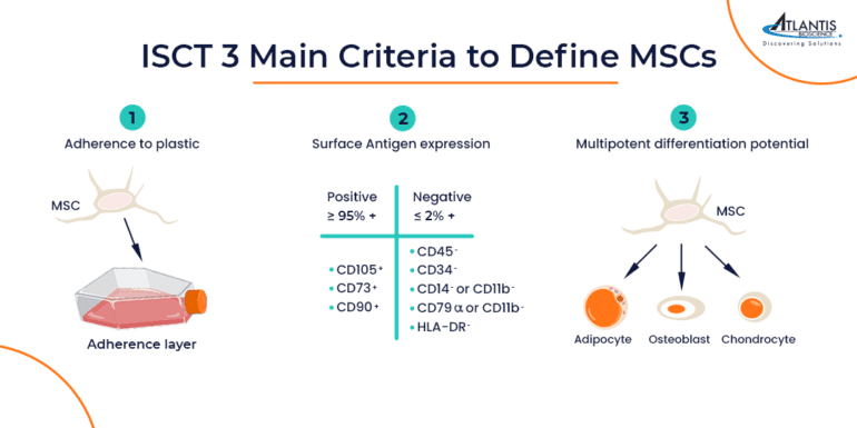
MSCs must adhere to plastic surfaces under standard cell culture conditions.
Multipotency:
They should demonstrate the capability to differentiate into osteoblasts, adipocytes, and chondroblasts in vitro.
Surface Antigen Expression:
MSCs must express specific surface antigens such as CD73, CD90, and CD105, while lacking the expression of lineage-specific markers including CD11b/CD14, CD19/CD79α, CD34, CD45, and HLA-DR.
MSC Culture Quality Control Challenges and Considerations
Heterogeneity and Variability
One of the major obstacles hindering the broader adoption of MSC-based therapies is the inherent heterogeneity of isolated cells. Variations in growth potential, differentiation capacity, and protein expression profiles have been observed across different tissue sources and production processes. This heterogeneity not only complicates the standardisation of MSC-based products but also impacts their therapeutic efficacy and safety profiles.
Clinical procedures utilise cell culture methodologies to amplify a limited population of primary MSCs obtained from specific tissue sources, expanding them through multiple passages to achieve a clinically significant cell count. The absence of universally accepted protocols for MSC isolation and expansion results in considerable variability in in vitro culture techniques across research entities. This variability complicates the comparability of findings across different studies.
Effective expansion strategies strive to enhance cell numbers substantially while preserving the therapeutic potential of MSCs. Notably, MSCs exhibit a decline in proliferation rates over time and with successive passages, eventually reaching a senescent state characterised by halted growth. Additionally, prolonged culture durations may compromise the cells’ differentiation capabilities.
Therefore, the establishment of standardised protocols, optimisation of culture conditions, and implementation of rigorous quality control measures are imperative to maintain consistent MSC attributes and functionalities across various batches.
MSC Culture Key Quality Control Parameters
Expression of cell surface antigens (CD markers)
The ISCT proposed a set of positive and negative markers that enable researchers to distinguish MSCs from other cells in the bone marrow compartment. The negative markers were selected to include surface antigens that are expressed by hematopoietic cells, while the positive markers were chosen for their absence from most hematopoietic cells.
Since the publication of the ISCT-proposed markers in 2006, several additional markers have gained widespread acceptance as MSC markers. This expanding panel of MSC-associated markers allows researchers to simultaneously verify the expression of multiple MSC-associated surface antigens, thereby increasing confidence in the identification and verification of isolated MSCs. Some widely used MSC markers include CD29, CD44, CD146, CD166, CD271, and STRO-1.
Marker expression analysis is typically performed using flow cytometry, which can reveal the phenotype of individual cells. Multicolour flow cytometry enables users to examine several markers simultaneously. Additionally, many flow cytometers are equipped with cell-sorting functions that can be used to enrich specific MSC populations and remove dead cells from the analysis.
Cell Viability and Proliferation
The viability of harvested MSCs can be assessed using trypan blue staining or 7-amino-actinomycin D (7-AAD) exclusion methods. To achieve optimal clinical outcomes with immediately applied MSCs and to ensure acceptable viability post-cryopreservation, the primary viability should exceed 90%.
The Colony-Forming Unit (CFU) assay serves as a widely recognised method for evaluating the self-renewal capacity of MSCs. This assay quantifies the efficiency with which MSCs generate colony units when seeded at clonogenic densities in monolayer culture on tissue culture plastic. Usually, single-cell suspensions of MSCs undergo serial dilution before being plated on culture plates. Upon becoming visible, typically around day 8 or 10, the colonies are stained with Giemsa and subsequently counted. Today, the CFU-F assay is employed in vitro to assess the quality of MSC preparations intended for both preclinical research and clinical trials.
Tri-Lineage Differentiation Potential
MSCs are characterised by their remarkable ability to differentiate into three distinct lineages: adipocytes, chondrocytes, and osteocytes when cultured under specific conditions. Successful differentiation can be verified by staining the differentiated cells for expression of lineage markers and/or by assessing the functionality of the cells. Confirmation of tri-lineage differentiation provides excellent evidence for the verification of MSC identity.
Osteogenic Differentiation:
MSCs can be directed to differentiate into osteoblasts, specialised cells responsible for bone formation and mineralisation. The osteogenic differentiation process involves the production of a mineralised extracellular matrix by osteoblasts. To assess osteogenic differentiation, researchers often measure the activity of specific markers such as alkaline phosphatase (ALP) or utilise staining techniques like Alizarin Red S to visualise calcium deposits, indicative of mature bone formation.
Adipogenic Differentiation:
When subjected to adipogenic induction conditions, MSCs can differentiate into adipocytes, cells specialised in lipid storage. The adipogenic differentiation process results in the accumulation of lipid droplets within the differentiated cells. To confirm adipogenic differentiation, Oil Red O staining is commonly employed, allowing for the visualisation of lipid accumulation and confirming the adipocyte phenotype.
Chondrogenic Differentiation:
MSCs possess the capability to differentiate into chondrocytes, the primary cells found in cartilage tissue responsible for producing cartilage-specific extracellular matrix components. Chondrogenic differentiation is crucial for cartilage repair and regeneration applications. Histological stains such as Alcian Blue are frequently used to detect the presence of glycosaminoglycans, confirming the formation of cartilage tissue and validating the chondrogenic differentiation potential of MSCs.
Functional Assays for Assessing MSC Potency and Therapeutic Potential
While the in vitro differentiation of MSCs into osteogenic, chondrogenic, and adipogenic lineages serves as a foundational characteristic, the clinical development of MSC-based therapies necessitates the establishment of potency assays. These assays aim to evaluate specific biological functional attributes that align with the anticipated clinical mechanisms of action of MSCs. Below are several key functional assays employed to assess MSC potency and therapeutic efficacy:
Immunomodulatory assays:
Understanding the immunomodulatory properties of MSCs is crucial, given their potential applications in immune-related disorders and transplantation. Co-culture assays involving immune cells, such as T cells, provide valuable insights into the immunosuppressive effects of MSCs, enabling researchers to evaluate their ability to modulate immune responses effectively.
Migration and homing assays:
MSCs possess inherent migratory capabilities, allowing them to home in on specific tissue injury sites or inflammatory microenvironments. Transwell migration assays or wound healing assays offer effective platforms for assessing MSC migration in response to various chemokines or stimuli, thereby elucidating their homing potential and tissue-targeting efficiency.
Angiogenic Assays:
The pro-angiogenic properties of MSCs hold significant promise for enhancing tissue regeneration and repair processes. In vitro tube formation assays, which involve co-culturing MSCs with endothelial cells, provide a robust platform for evaluating the angiogenic potential of MSCs and their capacity to stimulate new blood vessel formation, a critical step in tissue regeneration and wound healing.
Paracrine Activity Assays:
MSCs exert therapeutic effects through the secretion of various paracrine factors, including growth factors, cytokines, and extracellular vesicles. Quantitative assays such as enzyme-linked immunosorbent assays (ELISA) or multiplex cytokine assays facilitate the comprehensive analysis of MSC-secreted factors, offering insights into their paracrine activity and therapeutic potential.
Mechanical Properties:
Recent studies have also begun to explore the mechanical properties of MSCs, such as their stiffness and viscoelastic properties, which may influence their behaviour and functionality in vivo. Advanced techniques like atomic force microscopy (AFM) or rheological analyses provide valuable data on MSC mechanical properties, contributing to a more comprehensive understanding of their biological characteristics and therapeutic applications.
References
Andrzejewska A, Lukomska B, Janowski M. Concise Review: Mesenchymal Stem Cells: From Roots to Boost. Stem Cells. 2019;37(7):855-864. doi:10.1002/stem.3016
Dominici M, Le Blanc K, Mueller I, et al. Minimal criteria for defining multipotent mesenchymal stromal cells. The International Society for Cellular Therapy position statement. Cytotherapy. 2006;8(4):315-317. doi:10.1080/14653240600855905
Fonseca LN, Bolívar-Moná S, Agudelo T, et al. Cell surface markers for mesenchymal stem cells related to the skeletal system: A scoping review. Heliyon. 2023;9(2):e13464. Published 2023 Feb 10. doi:10.1016/j.heliyon.2023.e13464
Zhou T, Yuan Z, Weng J, Pei D, Du X, He C, Lai P. Challenges and advances in clinical applications of mesenchymal stromal cells. J Hematol Oncol. 2021 Feb 12;14(1):24. doi: 10.1186/s13045-021-01037-x
The Future is Flexible: Summary Substrate stiffness serves as a critical determinant of MSC behaviour, exerting profound effects on adhesion, proliferation, migration, and differentiation. Understanding
The use of mesenchymal stem cells (MSCs) in clinical trials has witnessed a global surge, positioning MSCs as one of the most sought-after regenerative cell
HOW CAN WE HELP YOU?Our specialists are to help you find the best product for your application. We will be happy to help you find the right product for the job.