RNA is often overshadowed by its more famous cousin, DNA. We traditionally think of RNA as a blueprint for proteins, carrying instructions from DNA to be translated into the building blocks of life. However, this isn’t the whole story. There’s a whole class of RNA molecules often referred to as the “silent regulators” – non-coding RNAs (ncRNAs).
Understanding Non-Coding RNAs
ncRNAs are a diverse class of RNA molecules that are transcribed from DNA but are not translated into proteins. Despite their lack of involvement in protein coding, ncRNAs constitute a substantial portion of the cellular RNA pool. In fact, they can comprise anywhere from 70% to 90% or more of the total RNA content in eukaryotic cells. This prevalence underscores their pivotal roles in cellular regulation and function, distinct from the messenger RNA (mRNA) responsible for protein synthesis.
Classification and Roles of ncRNA
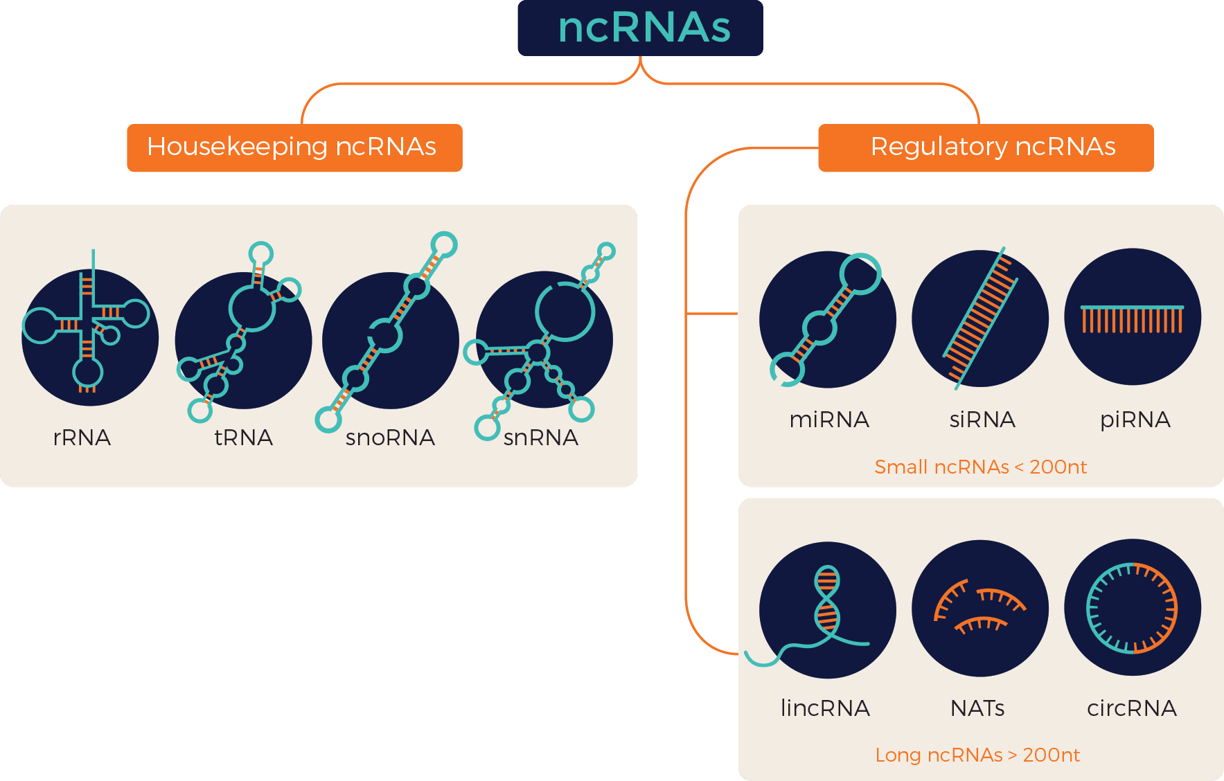
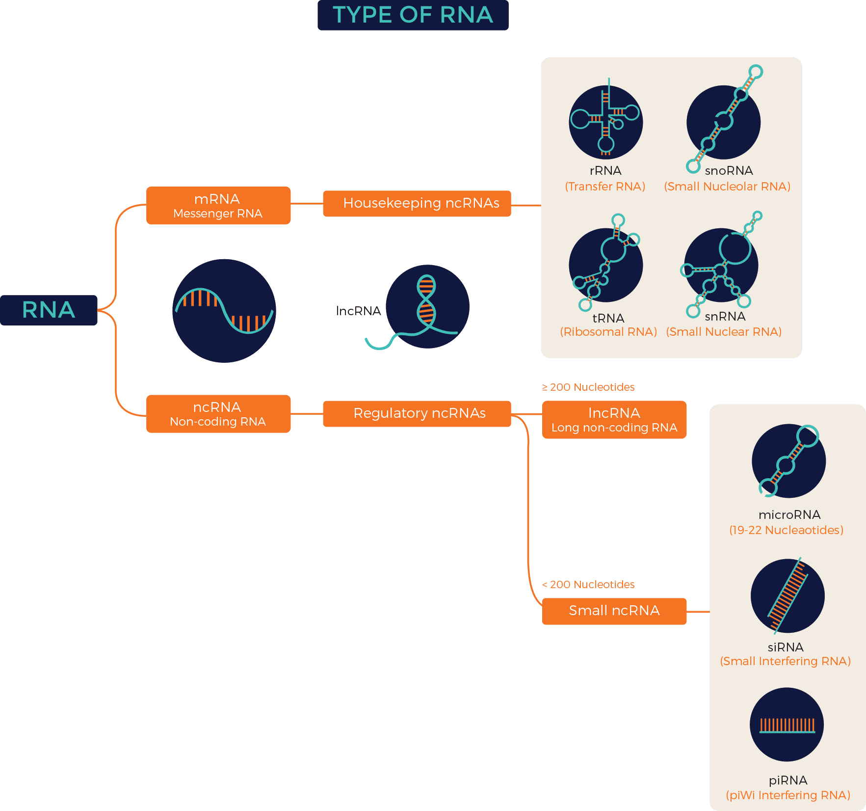
Figure 1: Types of ncRNAs
ncRNAs are broadly classified into two categories: housekeeping and regulatory RNAs, based on their biological roles.
Housekeeping ncRNAs:
Housekeeping ncRNAs are foundational components expressed abundantly and universally across cells. They primarily oversee essential cellular functions. Examples include:
Ribosomal RNAs (rRNAs) and Transfer RNAs (tRNAs): Constituting the majority of ncRNAs in mammalian cells, these molecules play indispensable roles in protein synthesis.
Small Nuclear RNAs (snRNAs) and Small Nucleolar RNAs (snoRNAs): Essential for intron splicing, RNA processing, and ribosomal RNA modification, snRNAs and snoRNAs contribute significantly to cellular maintenance. Due to their compact size, both are also categorised as small ncRNAs.
Regulatory ncRNAs:
In contrast to their housekeeping counterparts, regulatory ncRNAs exhibit remarkable diversity in expression and function. They are further classified into small ncRNAs (< 200 nt) and long ncRNAs (> 200 nt).
Small ncRNAs:
This category encompasses several classes, including:
MicroRNAs (miRNAs): These single-stranded molecules, typically 18-22 nucleotides long, modulate gene expression post-transcriptionally by targeting mRNA, leading to translational repression or degradation. miRNAs play pivotal roles in development, cell proliferation, differentiation, and disease pathogenesis.
Small Interfering RNAs (siRNAs): Double-stranded RNA molecules, approximately 20-25 nucleotides in length, siRNAs mediate RNA interference with high specificity, making them valuable tools for gene silencing and therapeutic interventions.
Piwi-interacting RNAs (piRNAs): These 24-31 nucleotide-long ncRNAs, unique to animals, regulate gene expression at both transcriptional and post-transcriptional levels, often through association with PIWI proteins.
Long Noncoding RNAs (lncRNAs):
With lengths exceeding 200 nucleotides, lncRNAs are usually folded into three-dimensional structures, and they boast diverse regulatory functions. Examples include:
Intergenic lincRNAs, Intronic ncRNAs, Sense and Antisense lncRNAs, bidirectional ncRNAs: These subclasses, delineated by genomic location, mechanism of action, or effect on DNA targets, exemplify the versatility of lncRNAs in modulating gene expression at transcriptional, post-transcriptional, and epigenetic levels.
Natural Antisense Transcripts (NATs): Complementary to other RNA molecules, NATs regulate gene expression and function by binding to sense RNA counterparts, influencing their activity.
Circular RNAs (circRNAs): With their closed-loop structures, circRNAs are highly stable and resist degradation and serve diverse roles – acting as microRNA sponges, RNA-binding protein sequesters, or regulators of transcription and splicing. CircRNAs contribute to a wide array of biological processes including cell proliferation, differentiation, aging, and the pathogenesis of various diseases, such as cancer and neurological disorders.
Exploring the Intricacies of miRNAs in Cancer Biology
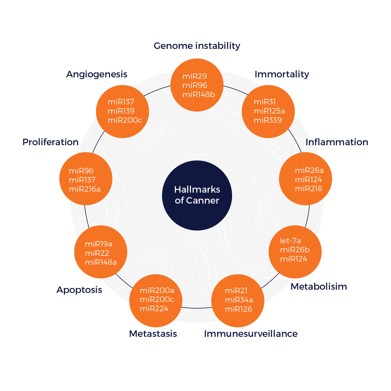
miRNAs, the most studied ncRNAs constitute a prevalent class of ncRNAs, intricately involved in the regulation of gene expression. Research has illuminated their pervasive role in numerous diseases, particularly cancer, where their dysregulation is a common occurrence. In the intricate landscape of cancer biology, miRNAs emerge as dual players, exhibiting both tumour-suppressive and oncogenic properties. Moreover, factors pivotal for miRNA biogenesis have been implicated in various cancers. This convergence of miRNA dysregulation and its associated biogenetic machinery underscores the profound impact of miRNAs on the initiation, progression, and therapeutic response of cancer. Notably, miRNAs have been intricately linked to the hallmarks of cancer, including sustained proliferation, evasion of growth suppressors, resistance to cell death, angiogenesis, invasion, and metastasis, further highlighting their multifaceted involvement in shaping the malignant phenotype.
Figure 2: Association between microRNAs and the hallmarks of cancer. Each hallmark shows three examples of microRNAs that influence the particular cellular function in certain types of cancer. Credit: Pichler M and Calin GA, doi: 10.1038/bjc.2015.253 Reproduced under the Creative Commons license
Roles of miRNAs in Cancer
Oncogenesis: miRNAs play pivotal roles in cancer development by functioning as either tumour suppressors or oncogenes. For instance, miR-34 acts as a tumour suppressor by downregulating genes involved in cell cycle progression, thereby inhibiting uncontrolled cell division, a hallmark of cancer. Conversely, oncogenic miRNAs like miR-21 promote tumorigenesis by targeting genes that regulate cell cycle inhibitors or apoptosis activators, fostering a pro-cancerous environment.
Metastasis: The spread of cancer to distant organs, known as metastasis, is a significant challenge in treatment. miRNAs significantly influence this process by regulating the epithelial-mesenchymal transition (EMT), a cellular transformation where epithelial cells acquire the migratory and invasive properties of mesenchymal cells. For example, miR-10b, which is controlled by an EMT facilitator, Twist1, promotes metastasis, with elevated expression seen in metastatic breast cancer.
Furthermore, researchers are exploring miRNA signatures—unique sets of miRNAs with potential to predict a tumour’s metastatic potential. Identifying such signatures could provide valuable insights into cancer aggressiveness and guide treatment decisions.
Biomarkers: Reliable cancer biomarkers are crucial, and miRNAs show promising potential. Their stability in bodily fluids makes them attractive targets for non-invasive detection methods. Investigating specific sets of miRNAs, such as miR10b, miR-135b, miR-196a, and miR-203, holds promise for early cancer detection and prognosis in various cancers.
Chemoresistance: Chemotherapy resistance can render treatment ineffective, and miRNAs play a key role in regulating the drug sensitivity of tumour cells. They can contribute to chemoresistance by various mechanisms, including influencing tumour stem cells, promoting angiogenesis, altering drug targets, and regulating drug resistance-related proteins. Understanding how miRNAs influence chemoresistance is crucial for identifying high-risk patients and developing strategies to overcome this challenge. Additionally, miRNA profiles might predict a patient’s likelihood of developing chemoresistance, enabling personalised treatment approaches.
Therapeutic Potential: miRNA-based therapies represent a transformative approach to cancer treatment. One strategy involves replacing downregulated tumour suppressor miRNAs using synthetic miRNA mimics to restore their function and suppress cancer cell growth. For instance, synthetic miR-34a analogs have shown anti-tumourigenic effects in murine models, although safety concerns led to the termination of a clinical trial. Another promising strategy utilises miRNA antagonists to block the function of oncogenic miRNAs. For example, anti-miR-10b has demonstrated suppression of breast cancer metastasis in preclinical studies. While miRNA-based therapies are in early stages, they hold significant potential for future cancer treatment.
Tools for Studying ncRNAs
The study of ncRNAs requires a diverse array of sophisticated tools and techniques tailored to isolate, characterise, and analyse these elusive molecules. Here, we delve into some of the key methodologies utilised by researchers in this field:
1. RNA Isolation: Isolating RNA is the foundational step in studying ncRNAs. Techniques such as phenol-chloroform extraction, column-based purification kits, and automated platforms enable researchers to extract high-quality total RNA from various sources, including cells, tissues, and biofluids. This isolation process is crucial for subsequent analyses, including quantitative PCR (qPCR), microarray analysis, and RNA sequencing.
2. Quantitative PCR (qPCR): Quantitative PCR is a cornerstone technique for quantifying the expression levels of ncRNAs with high specificity and sensitivity. Utilising fluorescently labelled probes or DNA binding dyes, qPCR enables the accurate measurement of ncRNA abundance in biological samples. Custom-designed primers targeting specific ncRNAs allow researchers to quantify their expression under different experimental conditions or disease states, providing valuable insights into their roles in cellular processes and pathologies.
3. RNA Sequencing: RNA sequencing (RNA-seq) has revolutionised the field of transcriptomics, allowing comprehensive profiling of ncRNA expression in a high-throughput manner. This next-generation sequencing technology enables the identification of known and novel ncRNAs, differential expression analysis, and characterisation of RNA isoforms. RNA-seq datasets provide deep insights into the complexity and dynamics of the ncRNA transcriptome, facilitating the discovery of regulatory networks and disease-associated signatures.
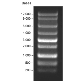
4. Northern Blotting: Northern blotting remains a classical technique for analysing RNA expression and size distribution. While less commonly used compared to qPCR and RNA-seq, Northern blotting offers advantages in detecting specific ncRNA species and assessing their abundance and molecular weight. By employing labelled probes complementary to target ncRNAs, Northern blotting provides qualitative and semi-quantitative information on ncRNA expression patterns across different experimental conditions or tissues.
5. Fluorescence In Situ Hybridisation (FISH): Fluorescence in situ hybridisation enables the visualization and localisation of ncRNAs within cells or tissues. By using fluorescently labelled probes complementary to specific ncRNA sequences, FISH allows researchers to spatially map ncRNA expression patterns and subcellular distribution. This technique provides valuable insights into the spatial regulation of ncRNAs and their potential roles in cellular organisation and function.
6. Bioinformatics Tools: Bioinformatics plays a critical role in analysing and interpreting ncRNA data generated from experimental assays. Computational tools and algorithms facilitate ncRNA discovery, prediction of ncRNA targets, functional annotation, and pathway analysis. These tools enable researchers to integrate diverse datasets, identify regulatory networks, and unravel the complex interplay between ncRNAs and cellular processes.
Conclusion
In conclusion, our exploration of RNA has revealed a far richer landscape than the simple messenger molecule initially described. NcRNAs, exemplified by miRNAs, demonstrate a remarkable capacity for regulating gene expression and influencing complex cellular processes. As discussed, miRNAs exhibit a fascinating duality in cancer biology, functioning as both tumour suppressors and oncogenes. Their potential as robust biomarkers for early cancer detection and personalised treatment strategies based on chemoresistance profiles holds immense promise for future clinical applications.
Unveiling the secrets of this intricate world requires specialised tools. Thankfully, researchers have developed a sophisticated arsenal of bioinformatics tools and wet-lab techniques specifically designed to study ncRNAs. These tools allow scientists to isolate, sequence, and analyse the expression patterns of these diverse molecules, providing deeper insights into their roles in health and disease.
Undoubtedly, further research into the intricacies of ncRNA function, facilitated by these ever-evolving tools, will continue to unveil their profound impact on human health and disease, paving the way for novel therapeutic interventions.
References
Bader AG. miR-34 – a microRNA replacement therapy is headed to the clinic. Front Genet. 2012;3:120. Published 2012 Jul 2. doi:10.3389/fgene.2012.00120
Good DJ. Non-Coding RNAs in Human Health and Diseases. Genes (Basel). 2023;14(7):1429. Published 2023 Jul 11. doi:10.3390/genes14071429
Kim, T., Croce, C.M. MicroRNA: trends in clinical trials of cancer diagnosis and therapy strategies. Exp Mol Med 55, 1314–1321 (2023). https://doi.org/10.1038/s12276-023-01050-9
Lekka E, Hall J. Noncoding RNAs in disease. FEBS Lett. 2018 Sep;592(17):2884-2900. doi: 10.1002/1873-3468.13182.
Li, H., Yang, B. Friend or foe: the role of microRNA in chemotherapy resistance. Acta Pharmacol Sin 34, 870–879 (2013). https://doi.org/10.1038/aps.2013.35
Ma L, Teruya-Feldstein J, Weinberg RA. Tumour invasion and metastasis initiated by microRNA-10b in breast cancer [published correction appears in Nature. 2008 Sep 11;455(7210):256]. Nature. 2007;449(7163):682-688. doi:10.1038/nature06174
Ma L, Reinhardt F, Pan E, Soutschek J, Bhat B, Marcusson EG, Teruya-Feldstein J, Bell GW, Weinberg RA. Therapeutic silencing of miR-10b inhibits metastasis in a mouse mammary tumor model. Nat Biotechnol. 2010 Apr;28(4):341-7. doi: 10.1038/nbt.1618.
Palazzo AF and Lee ES (2015) Non-coding RNA: what is functional and what is junk? Front. Genet. 6:2. doi: 10.3389/fgene.2015.00002
Pichler, M., Calin, G. MicroRNAs in cancer: from developmental genes in worms to their clinical application in patients. Br J Cancer 113, 569–573 (2015). https://doi.org/10.1038/bjc.2015.253
Wu S, Wu Y, Deng S, Lei X, Yang X. Emerging roles of noncoding RNAs in human cancers. Discov Oncol. 2023;14(1):128. Published 2023 Jul 13. doi:10.1007/s12672-023-00728-w
Zhang C, Sun C, Zhao Y, Wang Q, Guo J, Ye B, Yu G. Overview of MicroRNAs as Diagnostic and Prognostic Biomarkers for High-Incidence Cancers in 2021. Int J Mol Sci. 2022 Sep 27;23(19):11389. doi: 10.3390/ijms231911389.
Extracellular vesicles (EVs) are revolutionising our understanding of cell-to-cell communication. These ubiquitous messengers encased in a lipid bilayer membrane, are nano-sized packages actively released by
Exosomes are nano-sized extracellular vesicles ranging 30-150 nanometers. They are readily released by most cells and can be found in all body fluids including plasma, serum, saliva, cerebrospinal fluid, urine and cell culture media.
HOW CAN WE HELP YOU?Our specialists are to help you find the best product for your application. We will be happy to help you find the right product for the job.