- Your cart is empty
- Continue Shopping


Nanomedicines such as extracellular vesicles (EVs), liposomes, and lipid nanoparticles (LNPs) hold great potential for advancing medicine. In recent years, EVs have gained significant attention for their potential in diagnostics, therapeutics, and drug delivery. These nanoscale vesicles, secreted by cells, carry bioactive molecules such as proteins, lipids, and nucleic acids, making them ideal candidates for intercellular communication and biomarkers. Their natural origin and ability to deliver cargo to specific cells position EVs as promising tools in the clinical space, particularly in areas like cancer therapy, regenerative medicine, and vaccine delivery.
Despite their potential, translating EVs-based applications into clinical practice depends heavily on their reliable characterisation. To develop safe and effective EVs therapies, researchers must precisely understand their properties, such as size, surface charge, and molecular composition. Characterisation is essential not only for understanding their function but also for meeting stringent regulatory requirements.
In the following sections, we’ll explore the critical role of EVs characterisation in unlocking their clinical potential. We will compare common techniques, highlighting their strengths and limitations, and introduce Tunable Resistive Pulse Sensing (TRPS) as an emerging technology that is reshaping the field. Finally, we will introduce the Exoid, a cutting-edge instrument harnessing TRPS technology to deliver precise, reproducible, and comprehensive EV characterisation, setting a new benchmark for nanoparticle research and clinical applications.
To advance the clinical application of EVs, it is crucial to thoroughly evaluate specific critical quality attributes (CQAs) that influence their functionality and therapeutic potential. Below are the key parameters that must be assessed:
EVs are composed of diverse subtypes differing in size, composition, and biogenic properties. The characterisation of EV subtypes is complex. Particle size is a critical attribute of EVs that directly affects their stability, biodistribution, and biological activity. The size of EVs influences their ability to evade immune clearance, interact efficiently with target cells, and deliver therapeutic cargo to specific tissues. For instance, smaller EVs may circulate longer in the bloodstream, enhancing their delivery potential, while larger ones may be more prone to immune system recognition and clearance.
The particle size distribution (PSD) further reflects the uniformity of an EV population, which is crucial for achieving consistent biological behaviour. A narrow PSD indicates a homogeneous population, which is essential for reproducibility and ensures that the EVs exhibit predictable interactions and therapeutic effects. Conversely, a broad PSD may lead to variability in EVs function and reduce overall efficacy in clinical applications.
Accurate measurement of both particle size and PSD is vital for quality control and batch-to-batch consistency during production. In addition, understanding these parameters allows researchers to optimise EVs formulations for specific therapeutic goals, such as enhancing delivery efficiency or tailoring biodistribution. These metrics are also important for meeting regulatory standards, as variations in size and distribution can affect product safety and efficacy. Consequently, particle size and PSD analysis serve as foundational steps in the development, production, and clinical translation of EV-based nanomedicines.
The surface charge of EVs, quantified as zeta potential, is crucial for their stability and interactions within biological systems. A higher absolute zeta potential signifies stronger electrostatic repulsion between particles, minimising the risk of aggregation during storage or administration. In contrast, a lower absolute zeta potential indicates weaker repulsion, increasing the likelihood of particle aggregation.
By assessing zeta potential under varying preparation conditions, researchers can optimise EV formulations to enhance stability and functionality, such as improving cellular uptake and interaction. This parameter also serves as a valuable quality control metric during EV production and purification, ensuring consistent quality and minimising batch-to-batch variability. Consequently, measuring zeta potential is a key step in refining and standardising EV formulations for nanomedicine applications
The concentration of EVs in a sample is a crucial parameter that influences their therapeutic and diagnostic efficacy. Accurate measurement of EVs concentration ensures consistent quality across production batches, which is vital for reproducibility in both research and clinical applications. It also directly affects the amount of bioactive molecules delivered to target cells, determining the therapeutic outcome.
EVs concentration not only reflects the quantity of particles but also indicates the quality of the sample and the efficiency of isolation methods. A precise concentration measurement ensures that the isolation process has been effective and free from contaminants, which is essential for both the purity and functionality of the EVs preparation.
In clinical and diagnostic applications, maintaining the correct EVs concentration is crucial for achieving desired effects and reliable results. Insufficient concentration can reduce efficacy, while excessive concentration may pose safety risks. Moreover, concentration serves as a key quality control metric, ensuring batch-to-batch consistency and regulatory compliance for clinical use.
By precisely characterising these attributes, researchers can ensure the quality and performance of EVs-based products, paving the way for their successful integration into clinical applications.
Several techniques are available for the characterisation of EVs, each offering unique insights into their properties. These methods can be broadly categorised based on their ability to measure size, morphology, surface charge, and concentration, with some methods excelling in specific parameters while others provide a more comprehensive analysis.
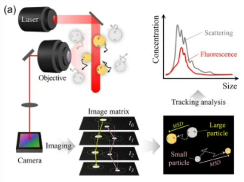
Nanoparticle Tracking Analysis (NTA) is a widely used technique for characterising EVs, offering real-time analysis of PSD and concentration. NTA works by visualising individual particles in suspension and tracking their Brownian motion under laser illumination. The motion is analysed to calculate the diffusion constant, which is then used in the Stokes-Einstein equation to determine the hydrodynamic diameter of particles. NTA also accounts for environmental factors such as temperature and viscosity, ensuring accurate size measurements.
NTA can also be used with fluorescence-based measurements, enabling researchers to examine labelled subpopulations of EVs. Particle concentration is calculated based on the number of tracked particles within the estimated illumination volume, providing quantitative insights into EV abundance.
Despite its advantages, NTA has limitations. It is less accurate for highly concentrated samples, requires careful calibration, and has a limited dynamic range (10⁶–10⁹ particles/mL). Large sample volumes (>250 µL) are typically required, and the technique is restricted to low-viscosity samples. Additionally, NTA requires a vibration-free environment to ensure reliable results.
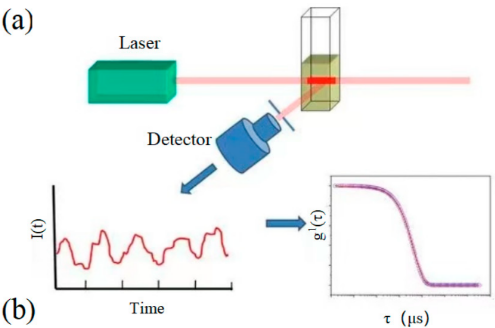
Dynamic Light Scattering (DLS) is another widely used technique for measuring the size distribution of EVs. By analysing the scattering of light due to the Brownian motion of particles, DLS estimates the hydrodynamic diameter of EVs in suspension. It is a top-down ensemble analysis method that calculates particle size from fluctuation signals across the sample.
While DLS is effective for determining the average hydrodynamic diameter, its sensitivity decreases with polydisperse populations, and it may overestimate the size of non-spherical EVs. DLS also struggles with very small or large particles, and since it’s not specific to EVs, it can detect co-isolated particles such as lipoproteins and protein aggregates, potentially confounding results.
An improved version, Multi-angle Dynamic Light Scattering (MADLS), addresses some of these limitations by measuring scattering at multiple angles. MADLS offers more accurate PSD and greater resolution, revealing populations that might be weakly scattered at a single detection angle.
However, DLS and MADLS are generally better suited for monodisperse samples ranging from 1 nm to 3 µm in diameter, with limitations for particles below 150 nm. For precise applications, such as the characterisation of EVs, accurate knowledge of optical properties (e.g., refractive index) is essential, as these can significantly influence results.
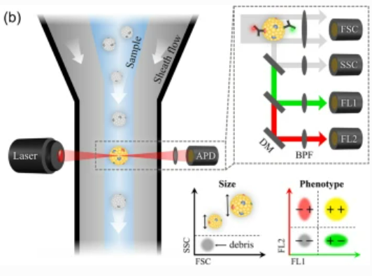
Nano Flow Cytometry (nFCM) is a specialised form of flow cytometry optimised for analysing nanoparticles, including EVs, with diameters of less than 1,000 nm. With the ability to detect EVs as small as 40 nm, nFCM provides high-resolution single-particle analysis.
In nFCM, particles are hydrodynamically focused into a central stream within a microchannel and exposed to laser excitation. The technique uses single-photon detectors (SPCMs) to measure light scattering and fluorescence signals. High-resolution side-scattered light (SSC) provides detailed information about particle size and concentration, while fluorescent labelling enables the identification of specific subpopulations.
However, nFCM has its limitations. At high particle concentrations, signal overlap can occur due to the “swarm effect,” leading to coincidental detection and compromised quantification accuracy. To mitigate this, samples need to be diluted to an optimal range (approximately 10⁷ to 10⁹ particles/mL). Additionally, amphiphilic dyes used for fluorescence labelling can form micelles or aggregates, potentially generating background signals. Therefore, it is essential to include controls, such as dye-only or buffer samples, to ensure the reliability of fluorescent labelling and subsequent analysis.
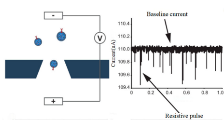
Tunable Resistive Pulse Sensing (TRPS) is a single-particle characterisation technique capable of measuring PSD, concentration, and zeta potential with high precision and accuracy. Unlike optical methods, TRPS operates electrically by detecting changes in impedance as particles traverse a nanopore. When a particle passes through the pore, the impedance change is recorded, allowing for the determination of particle size, concentration, and surface charge.
TRPS utilises a setup comprising two chambers separated by a nanoporous membrane with electrodes on either side. A potential difference across the membrane generates an electric current, and as a particle with low conductivity passes through the pore, resistance increases. This resistance results in a measurable drop in the current signal, from which the particle size and zeta potential can be calculated based on the pulse magnitude and duration. Concentration measurements are derived from the frequency of resistive pulses over time.
The tunability of TRPS, through adjustable pore sizes and multi-pressure calibration, enhances its accuracy for nanoparticle analysis. This feature allows for the precise measurement of EVs as small as ~37 nm, even in polydisperse samples. Additionally, surfactants can be employed to prevent particle aggregation, further improving the reliability of the measurements.
Compared to optical-based techniques like NTA, DLS, and nFCM, TRPS offers superior size resolution, particularly for monomodal and multimodal samples. Its higher sampling rate, reaching up to 500 kHz, outpaces the millisecond-scale exposure times required by optical methods, enabling faster and more dynamic analyses. Furthermore, TRPS can analyse particle concentrations across a broader range, from 10⁵ to 10¹⁴ particles/mL, making it particularly versatile for EV quantification. While TRPS excels in accuracy and dynamic range, proper optimisation of variables such as pore size, voltage, and sample preparation is essential to achieve reliable results.
Asymmetrical Flow Field-Flow Fractionation coupled with Multi-Angle Light Scattering (AF4-MALS) combines size-based separation with online detection to analyse nanoparticles. AF4 separates particles based on size by balancing their brownian motion and a perpendicular flow field within an open, unobstructed channel. Smaller particles elute earlier due to faster diffusion coefficients, while larger particles elute later.
Coupled with MALS, this method provides high-resolution particle size distribution, as well as mass- and number-weighted particle concentrations, with minimal sample interaction or deformation.
Centrifugal Liquid Sedimentation (CLS) measures the PSD based on sedimentation time in a rotating disc containing a density gradient, often of sucrose solution. According to Stokes’ Law, particles are separated by differences in size, density, and shape. Light-based detection methods are used to monitor sedimentation, with size calibrations typically relying on particle standards of known density. While CLS does not provide a direct number-weighted concentration output, it produces an intensity-based distribution indicative of mass-based concentration. Accurate concentration measurements may require additional knowledge of particle density, optical properties, and comparison with independent concentration methods.
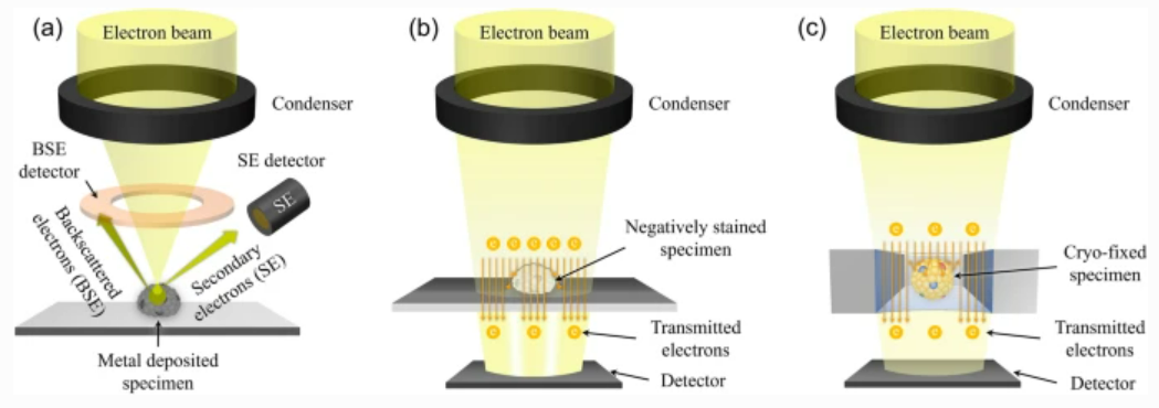
Electron microscopy (EM), encompassing transmission electron microscopy (TEM) and scanning electron microscopy (SEM), is widely used to image nanoscale samples, including EVs. TEM employs a beam of electrons transmitted through the sample, making it suitable for examining the internal structure of EVs. In contrast, SEM analyses scattered electrons, providing detailed surface structure information. Both techniques offer resolutions as fine as 1 nm.
Despite their high resolution, EM has limitations. Sample preparation for TEM and SEM involves fixation and dehydration, which can distort EV morphology. EVs lack internal structural support and may collapse into a cup shape upon dehydration. To address these issues, cryo-TEM can be employed. By embedding EV samples in vitreous ice at liquid nitrogen temperatures, cryo-TEM eliminates the need for fixation and dehydration, preserving the native morphology of EVs. However, challenges remain, such as potential perception bias from manually selected imaging areas and difficulty in estimating overall population characteristics.
Atomic Force Microscopy (AFM) is a powerful technique for capturing the surface structure of nanoscale samples. It operates by using a cantilever (probe) that interacts with the sample surface. As the probe deforms in response to surface morphological changes, the deformation is detected via laser reflection onto position-sensitive photodiodes. This enables AFM to generate true 3D images of surface topology, making it a valuable tool for surface structure analysis.
However, EVs lack internal structural support, which can lead to deformation during sample preparation and imaging. To enhance imaging quality, AFM is often combined with monoclonal antibody immobilisation, which stabilises the EVs for more accurate probe scans. Despite these challenges, AFM remains a reliable method for detailed surface topology determination.
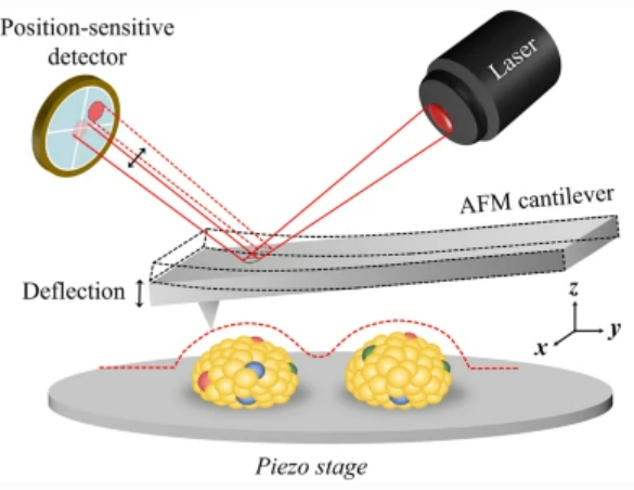
Table 1: Comparison of eight orthogonal methods for EVs characterisation.
| Single particle analysis | Ensemble analysis | |||||||
| Types | TRPS | NTA | nFCM | AFM | EM | AF4 MALS | CLS | MADLS |
| Detection Type | Electric current | Light scattering, fluorescence | Light scattering, fluorescence | Light position | Scattered/Transmitted electron | Light scattering | Light scattering | Light scattering |
| Detection Size | > 40 nm | > 50 nm | > 50 nm | > 1 nm | > 1–2 nm | > 20nm | > 20nm | > 1 nm |
| Concentration | Yes | Yes | Yes | No | No | Yes, not direct | Yes, not direct | Yes, not direct |
| Size | Yes | Yes | Yes | Yes | Yes | Yes | Yes | Yes |
| Charge | Yes | Yes | No | No | No | No | No | No |
| Shape | No | No | No | Yes | Yes | No | No | No |
Each analytical technique for nanoparticle characterisation has its own limitations. To fully characterise EVs—including parameters like PSD, concentration, and morphology—researchers often need to compare results from multiple orthogonal methods. This becomes especially critical when dealing with complex biological samples that exhibit a polydisperse distribution across a wide size range.
Precision and reliability are vital in nanoparticle research to produce high-quality data. Selecting the appropriate analytical technique is essential, particularly for polydisperse or intricate samples. While traditional methods such as DLS and NTA are widely used, advanced technologies like TRPS have emerged as powerful tools for precise, single-particle analysis.
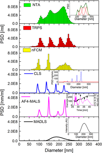
A comparative study by Vogel et al. (2021) highlighted the strengths of TRPS in overcoming challenges posed by complex samples. In tests with bimodal mixtures of NIST-traceable polystyrene particles (1:1 ratio of 100 and 200 nm), MADLS failed to resolve distinct particle features, showing only broad distributions. For trimodal mixtures (60, 100, and 150 nm particles), TRPS and nFCM delivered accurate resolution, while NTA underestimated the smallest particle population, reporting 2–5% instead of the expected 33.3%. Quadrimodal samples revealed further limitations in other techniques. While TRPS, CLS, and AF4-MALS achieved clear resolution, TRPS demonstrated strong repeatability across multiple runs. Although nFCM initially resolved quadrimodal samples, resolution diminished when data from multiple runs were averaged, merging peaks of 200 and 240 nm particles.
(CPN100/CPN150/CPN200/CPN240 at 25/25/25/25)
(CPN100/CPN150/CPN200/CPN240 at 10/50/30/10)
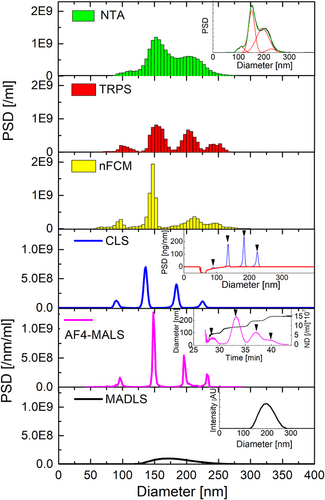
Figure 7: NTA, TRPS, nFCM, CLS, AF4-MALS and MADLS measurements of quadrimodal sample. Left: (CPN100/CPN150/CPN200/CPN240 at 25/25/25/25). Right: (CPN100/CPN150/CPN200/CPN240 at 10/50/30/10). NTA, TRPS, nFCM and MADLS measurements were averaged over 3 runs and CLS over 2. AF4-MALS was only performed once. Reference: https://doi.org/10.1002/jev2.12052
When analysing EV-enriched plasma fractions prepared via size exclusion chromatography, TRPS again excelled, providing repeatable size and concentration measurements with a coefficient of variation below 10%. By contrast, NTA consistently overestimated concentrations, and nFCM underestimated concentrations for smaller particles. AF4-MALS, while precise in certain contexts, struggled to quantify EV concentrations due to difficulties in adapting protocols for non-uniform biological samples.
As nanoparticle research advances into clinical and environmental applications, reliable techniques for measuring size, concentration, and surface charge are increasingly critical. The comparative study by Vogel et al. underscores the limitations of optical-based methods like NTA and MADLS, particularly for polydisperse or complex samples. In contrast, TRPS emerged as a standout method, offering high-resolution, reproducible results across a wide range of particle sizes and sample complexities. Its ability to resolve complex polydisperse samples and deliver high-resolution data makes TRPS a valuable tool in EV characterisation.
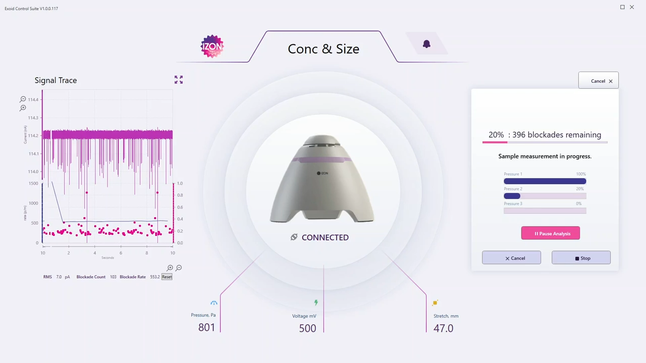
The Exoid is Izon’s state-of-the-art instrument for nanoparticle characterisation, employing TRPS—a cutting-edge technology unique to Izon. While Resistive Pulse Sensing (RPS) methods exist, the Exoid stands out as the only system with tunable capabilities, allowing unparalleled precision in particle size, concentration, and zeta potential measurements.
TRPS works by measuring individual nanoparticles as they pass through a tunable nanopore. By adjusting the nanopore size, users can tailor measurements to specific size ranges, ensuring high-resolution data. This single-particle approach avoids the biases inherent in ensemble techniques, where larger particles can obscure the size distribution of smaller populations. The ability to simultaneously measure particle size and zeta potential is particularly valuable for understanding the properties of complex particle dispersions, such as exosomes, lipid nanoparticles, and virus-like particles.
Accurate characterisation is critical for advancing EV applications in diagnostics and therapeutics. While traditional methods like DLS and NTA have limitations with complex, polydisperse samples, TRPS offers a transformative solution through precise, single-particle analysis. The Exoid, powered by TRPS, delivers unmatched accuracy for measuring size, concentration, and zeta potential, even for intricate samples. Its tunable nanopore technology, real-time calibration, and adaptability make it a valuable tool for EV research, ensuring high-resolution data that meets the demands of clinical and translational applications.
To learn how the Exoid can enhance your EV characterisation workflow, contact us today and take the next step in achieving precise and reliable results!
References
Kwon, Y., Park, J. Methods to analyze extracellular vesicles at single particle level. Micro and Nano Syst Lett 10, 14 (2022). https://doi.org/10.1186/s40486-022-00156-5
Marques, C.; Maurizi, L.; Borchard, G.; Jordan, O. Characterization Challenges of Self-Assembled Polymer-SPIONs Nanoparticles: Benefits of Orthogonal Methods. Int. J. Mol. Sci. 2022, 23, 16124. https://doi.org/10.3390/ijms232416124
Théry C, Witwer KW, Aikawa E, Alcaraz MJ, et. al., Minimal information for studies of extracellular vesicles 2018 (MISEV2018): a position statement of the International Society for Extracellular Vesicles and update of the MISEV2014 guidelines. J Extracell Vesicles. 2018 Nov 23;7(1):1535750. doi: 10.1080/20013078.2018.1535750.
Vogel R, Savage J, Muzard J, Camera GD, Vella G, Law A, Marchioni M, Mehn D, Geiss O, Peacock B, Aubert D, Calzolai L, Caputo F, Prina-Mello A. Measuring particle concentration of multimodal synthetic reference materials and extracellular vesicles with orthogonal techniques: Who is up to the challenge? J Extracell Vesicles. 2021 Jan;10(3):e12052. doi: 10.1002/jev2.12052.
Welsh JA, Goberdhan DCI, O’Driscoll L, Buzas EI, et. al., MISEV Consortium; Théry C, Witwer KW. Minimal information for studies of extracellular vesicles (MISEV2023): From basic to advanced approaches. J Extracell Vesicles. 2024 Feb;13(2):e12404. doi: 10.1002/jev2.12404.
Wu S, Zhao Y, Zhang Z, Zuo C, Wu H, Liu Y. The Advances and Applications of Characterization Technique for Exosomes: From Dynamic Light Scattering to Super-Resolution Imaging Technology. Photonics. 2024; 11(2):101. https://doi.org/10.3390/photonics11020101
Zhao Z, Wijerathne H, Godwin AK, Soper SA. Isolation and analysis methods of extracellular vesicles (EVs). Extracell Vesicles Circ Nucleic Acids. 2021;2:80-103. http://dx.doi.org/10.20517/evcna.2021.07

Connect With Our Technical Specialist.

Request For A Quotaiton
DON’T MISS OUR.
FOLLOW US ON SOCIAL MEDIA!
![]()
![]()
![]()
Contact our Customer Care, Sales & Scientific Assistance
Consult and asked questions about our products & services
Documentation of Technical & Safety Data Sheet, Guides and more..