Extracellular vesicles (EVs) have emerged as critical players in diagnostics, vaccine development, and therapeutic applications, driving the need for precise characterization tools. Exoid, powered by tunable resistive pulse sensing (TRPS), has become an essential instrument for researchers seeking accurate size distribution, concentration, and zeta potential measurements of EVs. Across various disciplines, research groups are leveraging Exoid to refine EV-based diagnostics, optimize vaccine formulations, and advance therapeutic applications. This blog highlights how different teams utilize Exoid for their EV characterization needs, demonstrating its impact on cutting-edge scientific advancements.
Using Exoid For Diagnostic Applications
Exosomal CD44 in BALF: A Promising Biomarker for Fibrotic Lung Diseases
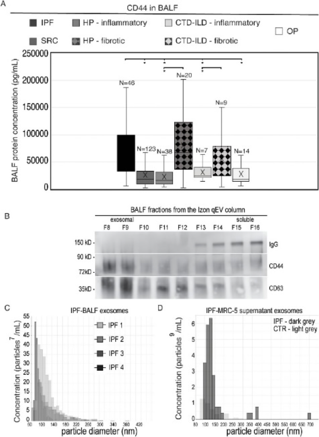
Diffuse parenchymal lung diseases (DPLDs), including idiopathic pulmonary fibrosis (IPF), are marked by lung inflammation and fibrosis, leading to progressive functional decline. Despite advancements, specific biomarkers for early fibrosis detection remain elusive. A recent study by Suchankova et al. identified exosomal CD44 in bronchoalveolar lavage fluid (BALF) as a potential diagnostic biomarker for pulmonary fibrosis, distinguishing fibrotic from non-fibrotic DPLDs.
Analysis of BALF from 257 DPLD patients revealed elevated CD44 levels in fibrotic DPLDs (IPF, fibrotic hypersensitivity pneumonitis, and fibrotic connective tissue disease-associated ILD). The exosomal origin of CD44 was confirmed using Izon qEV columns for isolation and characterized with the Exoid TRPS instrument using NP150 nanopores. CD44 effectively differentiated fibrotic from non-fibrotic DPLDs and correlated with high-resolution computer tomography (HRCT) imaging fibrosis patterns.
Figure 1. CD44 Evaluation in BALF from DPLD Patients. (A) CD44 levels in BALF analyzed by ELISA. (B) IPF-BALF fractions obtained via Izon qEV size-exclusion chromatography (SEC) were analyzed by Western blot for CD44, CD63 (exosomal marker), and IgG. (C, D) TRPS analysis of EV size (x-axis) and concentration (y-axis). Exosomal fractions from IPF-BALF (C) and conditioned medium of IPF-BALF-activated (IPF) or PBS-treated (CTR) MRC-5 cells (D) showed vesicle diameters (~150 nm), suggesting similar exosomal characteristics between in vivo BALF and in vitro fibroblast-derived exosomes.
Reference: doi: 10.3389/fimmu.2024.1479458 Reproduced under the Creative Commons licenseTable 1: Exosome Analysis by Exoid-TRPS Mean/mode particle diameter and concentration of exosomes in BALF from IPF patients (N = 4) and conditioned supernatants of BALF-activated MRC-5 cells (N = 3), measured using Nanopore 150 (range: 60–640 nm).
The findings highlight the potential of BALF-CD44 as a diagnostic biomarker for pulmonary fibrosis, offering a new approach to differentiate fibrotic from non-fibrotic DPLDs. This could lead to more accurate diagnoses, better patient stratification, and improved therapeutic outcomes in fibrotic lung diseases.
Plasma EV MicroRNA Signatures as Diagnostic Biomarkers for Melanoma
Melanoma is a highly aggressive skin cancer, accounting for over 80% of skin cancer-related deaths despite constituting only 1.8% of cases. While targeted therapies and immune checkpoint inhibitors have improved patient outcomes, metastatic melanoma remains a significant clinical challenge, with nearly 50% of advanced-stage patients succumbing within five years. Given the urgent need for non-invasive biomarkers, researchers have explored plasma EV (pEV)-associated microRNAs as potential diagnostic tools.
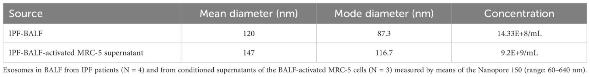
In this study, pEVs were isolated from plasma samples of melanoma patients and healthy donors. The particle size distribution and concentration of the purified EVs were determined using the Exoid TRPS measurement system. For each sample, an average of 500 particles was counted, and the data were compared with size and concentration reference calibration particles using a 150 nm nanopore. TRPS measurements of the pEVs from healthy donors revealed a particle concentration of 1.77e+09 particles/ml and an average particle diameter of 114 nm (standard deviation of 39.4 nm). In contrast, pEVs isolated from melanoma patients showed a higher particle concentration (2.89e+11 particles/ml) and a smaller average particle diameter of 98 nm (standard deviation of 11.7 nm).
Figure 2: Characterization of pEVs in Melanoma Patients and Healthy Controls. TEM visualization of EVs isolated from plasma, showing round vesicles (100-400 nm in diameter). Scale bar = 200 nm. (B) Western Blot analysis of exosomal markers (HSP70, TSG101, CD63, CD81) and calnexin in WM793 melanoma cells and EVs from control plasma. (C, D) Size distribution and concentration of pEVs from healthy donors (C) and melanoma patients (D) using TRPS.
Using a panel of 754 microRNAs, researchers identified 65 differentially expressed pEV-microRNAs in melanoma patients, with 44 upregulated and 21 downregulated. Functional analysis linked these microRNAs to critical biological processes such as angiogenesis, cell motility, and oncogenic signalling pathways.
Through LASSO logistic regression, a four-microRNA signature (miR-412-3p, miR-507, miR-1203, and miR-362-3p) was identified, demonstrating high diagnostic accuracy. This signature was successfully validated in independent cohorts, achieving an accuracy of 0.81 and an AUC of 0.94–0.96, reinforcing its robustness.
Key Findings and Implications
The findings highlight the potential of pEV-microRNA signatures as reliable liquid biopsy biomarkers for melanoma detection, paving the way for improved early diagnosis and disease monitoring.
Using Exoid For Vaccine Applications
Exploring Actinobacillus Pleuropneumoniae-Derived EVs for Vaccine Development
This study investigates the potential of Actinobacillus pleuropneumoniae-derived EVs (APP-EVs) as a vaccine candidate for combating APP infections in swine. The research compares APP-EVs to the commercial Coglapix vaccine, highlighting their immunogenicity and efficacy.
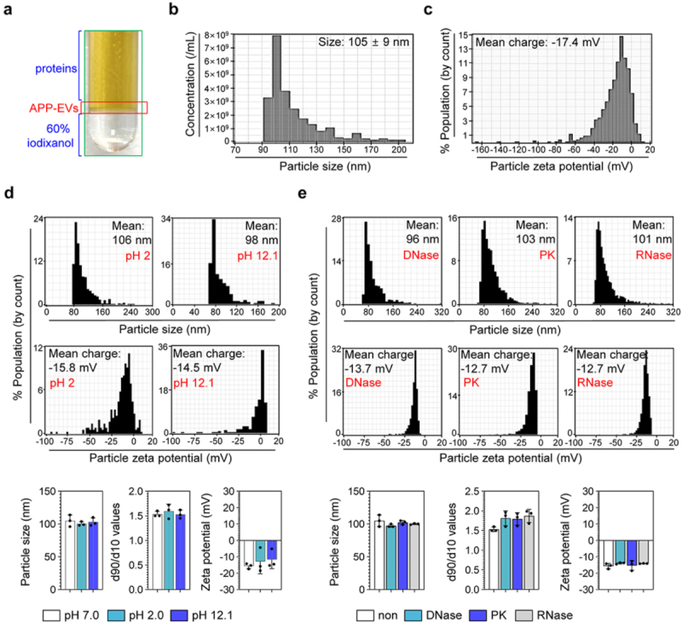
TRPS analysis using Exoid revealed that high-purity APP-EVs, isolated via TFF and ultracentrifugation, had an average size of 105 nm ± 9 nm and a stable zeta potential of −17.4 mV. Remarkably, APP-EVs maintained their size, distribution, and zeta potential under various stresses, including alkaline (pH 12.1) and acidic (pH 2.0) conditions, as well as exposure to DNase, RNase, and Proteinase K, demonstrating their stability.
Figure 3: Characterization and Stability of APP-EVs. APP-EVs were isolated using cushioned ultracentrifugation. The size (B) and zeta potential (C) of APP-EVs were analyzed with Exoid. The effects of pH changes (D; pH 2.0 and pH 12.1) and enzyme exposures (E; DNase, RNase, and Proteinase K) on size, zeta potential, and size distribution were also assessed using Exoid. Measurements were performed in triplicate, with histograms representing typical results.
These vesicles also contained a variety of metabolites, including amino acids, lipids, and nucleosides, contributing to their biological functions. When tested on dendritic cells (DCs), APP-EVs induced DC maturation through a Toll-like receptor 4 (TLR4)-dependent mechanism, promoting the production of pro-inflammatory cytokines such as IL-12p70. Importantly, APP-EVs did not cause cytotoxicity in DCs. In animal models, APP-EV immunization triggered robust Th1-mediated IgG responses, enhanced macrophage phagocytosis, and improved survival rates against APP infection. The study demonstrated that, unlike Coglapix, which induced stronger Th2 responses and notable toxicity, APP-EVs were safer and more effective in eliciting a Th1-dominant immune response.
Key Findings and Implications
In conclusion, APP-EVs show promise as a novel vaccine platform, capable of inducing potent and targeted T-cell responses, such as Th1, Th17, and cytotoxic T lymphocytes (CTLs), thereby offering a safer and more effective alternative to current vaccines for APP infection.
Isolation and Characterization of Spirulina Extracellular Vesicles (SPEVs): Potential as an Immunomodulatory Adjuvant
This study provides the first detailed analysis of EVs from Spirulina, a cyanobacterium recognized for its health benefits. The research isolates and characterizes these Spirulina-derived EVs (SPEVs), revealing their potential as immune modulators and adjuvants for vaccines. By using techniques like ultracentrifugation, Izon qEVsize-exclusion chromatography (SEC), and cryo-transmission electron microscopy (cryo-TEM), the study visualizes SPEVs as a combination of outer membrane vesicles (OMVs) and outer-inner membrane vesicles (OIMVs).
The immune response to SPEVs was assessed in a mouse model, where they were shown to significantly increase neutrophils and M1 macrophages, indicating a pro-inflammatory effect without causing toxicity or hypersensitivity. Additionally, SPEVs enhanced antigen-specific IgG responses more than 100-fold compared to an unadjuvanted vaccine, highlighting their potential as vaccine adjuvants.
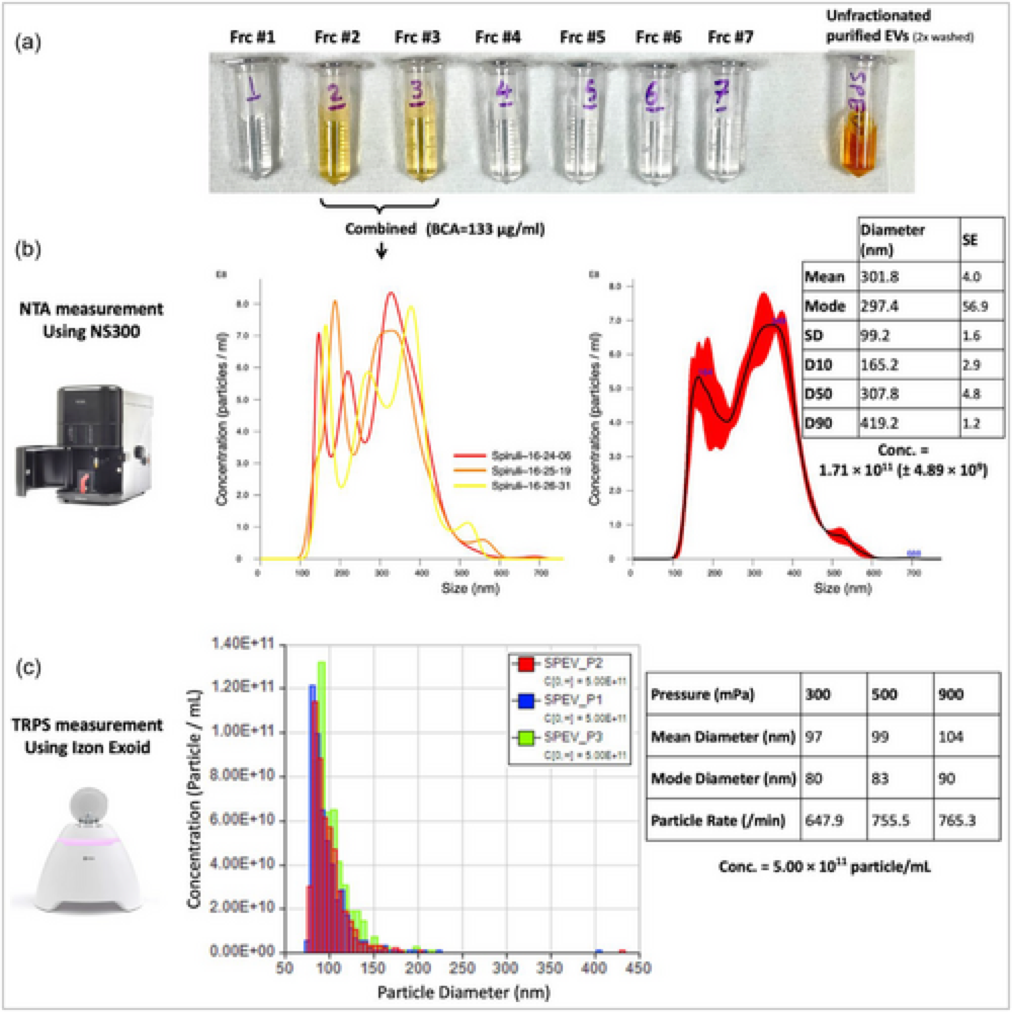
The study also reports differences in size distribution and concentration between two particle characterization methods: Nanoparticle Tracking Analysis (NTA) and TRPS. TRPS demonstrated a higher concentration of EVs (5 × 10¹¹ particles/mL) and a more consistent size range (75–225 nm, with a mean of 100 nm), while NTA reported a bimodal distribution with sizes up to 500 nm. Cryo-TEM confirmed the presence of EVs in the 100–200 nm range, consistent with TRPS results.
Notably, the study emphasizes the advantages of TRPS over NTA for accurately characterizing the concentration and size of SPEVs. TRPS provides more detailed and consistent results, making it a better technique for studying SPEVs and potentially other microbial EVs.
Figure 4: Analysis of the size distribution and concentration of isolated SPEVs using two different technologies. (A) Unfractionated SPEV suspension compared to fractionated EVs (fractions 1–7), and (B) Results from NTA and (C) TRPS. Reference: doi: 10.1002/jex2.70025 Reproduced under the Creative Commons license
Proteomic analysis identified 54 proteins in SPEVs, including several related to immune modulation, like carbohydrate-selective porins and S-layer homology proteins. These proteins could play a role in the observed pro-inflammatory effects. The study suggests that these properties could make SPEVs a valuable addition to the arsenal of natural adjuvants for vaccine development.
Key Findings and Implications
This study highlights the immunomodulatory potential of SPEVs as natural adjuvants to enhance vaccine efficacy. SPEVs activate both innate and adaptive immunity, improving antigen presentation and cytokine production. These findings suggest SPEVs as a promising alternative to synthetic adjuvants, offering a more sustainable and biocompatible approach to vaccine development.
Using Exoid For Therapeutic Applications
MSC-Derived EVs Promote Angiogenesis and Lymphangiogenesis in Ischemic Tissues via miRNAs and Proteins
Mesenchymal stem cell-derived EVs (MSC-EVs) play a crucial role in ischemic tissue repair by promoting angiogenesis and restoring blood flow. This study investigated the biological effects of bone marrow MSC-EVs on endothelial cells in both in vitro and in vivo ischemic models, highlighting their molecular composition and mechanisms of action. Proteomic and microRNA analyses revealed that MSC-EVs are enriched in bioactive molecules that regulate key regenerative pathways, including integrin α5 (Itgα5) and neuropilin-1 (NRP1), which are involved in lymphangiogenesis. Functional assays demonstrated that MSC-EVs enhance capillary-like tube formation, protect endothelial cells from apoptosis, and stimulate nitric oxide (NO) production via endothelial NO synthase activation. Importantly, in a murine hind limb ischemia model, MSC-EVs improved blood flow recovery and increased vascular density, reinforcing their therapeutic potential in ischemic diseases.
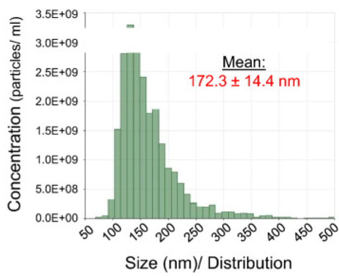
To characterize the MSC-EV preparations, TRPS was employed to analyze the size distribution of nanoparticles. Using an NP200 nanopore (analytical range 85–500 nm), samples were measured for 5 minutes or until 1000 particles were counted. Following ISEV recommendations for EV characterization, the study confirmed that the murine bone marrow MSC-EV isolates represented a heterogeneous population of particles, ranging from 70 to 430 nm in size, with a mean diameter of 172.3 ± 14.4 nm. These findings emphasize the variability in EV populations and underscore the importance of precise nanoparticle analysis to ensure consistency and reproducibility in therapeutic applications.
Figure 5: Representative histogram of particle size distribution in MSC-EVs specimen by IZON qNano.
These findings have important implications for regenerative medicine, particularly in developing cell-free therapies for ischemic conditions. By identifying key molecular players like Itgα5 and NRP1, this study supports targeted EV-based strategies for vascular and lymphatic regeneration. MSC-EVs’ roles in promoting angiogenesis and lymphangiogenesis suggest they could improve outcomes in conditions such as stroke, myocardial infarction, and peripheral artery disease.
Comparative Efficacy of Exosomes Derived from iMSCs and SMMSCs in Osteoarthritis Treatment
Osteoarthritis (OA) is a prevalent joint disease, affecting 10% of men and 18% of women over 60, leading to a significant healthcare burden. The treatment of OA has been challenging due to the lack of blood supply to cartilage and the limited regenerative capacity of chondrocytes. While MSCs from sources like bone marrow and adipose tissue have shown promise for OA treatment, issues such as tumour formation, ethical concerns, and immune rejection persist. Exosomes, secreted by MSCs, have emerged as a potential solution for tissue repair, as they mediate many of the therapeutic effects attributed to MSCs. However, the effectiveness of MSC-derived exosomes for OA treatment, particularly from synovial membrane-derived MSCs (SMMSCs) and induced pluripotent stem cell-derived MSCs (iMSCs), remains unstudied.
This study compared the therapeutic effects of exosomes from SMMSCs and iMSCs on OA treatment. The results demonstrated that exosomes from iMSCs (iMSC-Exos) had a superior therapeutic effect compared to SMMSC-derived exosomes (SMMSC-Exos) in a collagenase-induced mouse OA model. Histological analysis showed that iMSC-Exos promoted the repair of damaged cartilage, presenting hyaline features similar to normal cartilage, and significantly increased chondrocyte migration and proliferation in vitro. In contrast, SMMSC-Exos showed only moderate improvement, with weaker collagen II expression in treated cartilage.
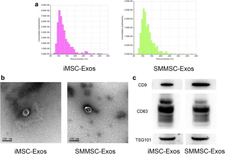
The exosomes derived from both iMSCs and SMMSCs were characterized to understand their properties better. TRPS analysis revealed that the majority of iMSC-Exos and SMMSC-Exos had sizes ranging from approximately 50 to 150 nm. TEM further confirmed that both exosome types exhibited a cup-shaped or round morphology with diameters between 50 and 200 nm.
Figure 6: Characterization of iMSC-Exos and SMMSC-Exos. (A) TRPS measurement showing exosome concentration and size distribution. (B) Morphological analysis of exosomes via transmission electron microscopy. (C) Western blot analysis of exosome-specific markers CD9, CD63, and TSG101.
This study suggests that iMSC-Exos may be more effective than SMMSC-Exos for OA treatment, offering superior therapeutic effects in both animal models and in vitro. Additionally, iMSCs present several advantages over SMMSCs, including non-invasive harvesting, the potential to eliminate immunosuppression, and an inexhaustible supply of autologous MSCs. These findings highlight the potential of iMSC-Exos as a novel, patient-specific, and scalable therapeutic approach for OA, with further research needed to explore the underlying mechanisms and optimize their clinical application.
References:
Hyun Park S, Kim YH, Lee HJ, Han JM, Seo BJ, Park GS, Kim C, Ryu YB, Kim WS. Immunogenicity and vaccine efficacy of Actinobacillus pleuropneumoniae-derived extracellular vesicles as a novel vaccine candidate. Virulence. 2025 Dec;16(1):2453818. doi: 10.1080/21505594.2025.2453818
Łabędź-Masłowska A, Vergori L, Kędracka-Krok S, Karnas E, Bobis-Wozowicz S, Sekuła-Stryjewska M, Sarna M, Andriantsitohaina R, Zuba-Surma EK. Mesenchymal stem cell-derived extracellular vesicles exert pro-angiogenic and pro-lymphangiogenic effects in ischemic tissues by transferring various microRNAs and proteins including ITGa5 and NRP1. J Nanobiotechnology. 2024 Feb 12;22(1):60. doi: 10.1186/s12951-024-02304-y.
Sabato C, Noviello TMR, Covre A, Coral S, Caruso FP, Besharat ZM, Splendiani E, Masuelli L, Battistelli C, Vacca A, Catanzaro G, Po A, Anichini A, Maio M, Ceccarelli M, Di Giacomo AM, Ferretti E. A novel microRNA signature for the detection of melanoma by liquid biopsy. J Transl Med. 2022 Oct 15;20(1):469. doi: 10.1186/s12967-022-03668-1.
Sharifpour MF, Sikder S, Wong Y, Koifman N, Thomas T, Courtney R, Seymour J, Loukas A. Characterization of Spirulina-derived extracellular vesicles and their potential as a vaccine adjuvant. J Extracell Biol. 2024 Dec 12;3(12):e70025. doi: 10.1002/jex2.70025.
Suchankova M, Zsemlye E, Urban J, Baráth P, Kohútová L, Siváková B, Ganovska M, Tibenska E, Szaboova K, Tedlova E, Juskanic D, Kluckova K, Kardohelyova M, Moskalets T, Ohradanova-Repic A, Babulic P, Bucova M, Leksa V. The bronchoalveolar lavage fluid CD44 as a marker for pulmonary fibrosis in diffuse parenchymal lung diseases. Front Immunol. 2025 Jan 13;15:1479458. doi: 10.3389/fimmu.2024.1479458.
Zhu Y, Wang Y, Zhao B, Niu X, Hu B, Li Q, Zhang J, Ding J, Chen Y, Wang Y. Comparison of exosomes secreted by induced pluripotent stem cell-derived mesenchymal stem cells and synovial membrane-derived mesenchymal stem cells for the treatment of osteoarthritis. Stem Cell Res Ther. 2017 Mar 9;8(1):64. doi: 10.1186/s13287-017-0510-9.
Cryopreservation is a technique designed to preserve the viability and functionality of biological specimens, such as cells, tissues, and organs, by cooling them to subzero
The concept of Critical Quality Attributes (CQAs) plays a pivotal role in a drug development process. According to the ICH Guideline Q8(R2) on Pharmaceutical Development,
Cell and gene therapies (CGTs), particularly autologous therapies like CAR-T/TCR-T cells, represent a breakthrough in personalized medicine. These treatments offer life-changing outcomes for patients with
HOW CAN WE HELP YOU?Our specialists are to help you find the best product for your application. We will be happy to help you find the right product for the job.