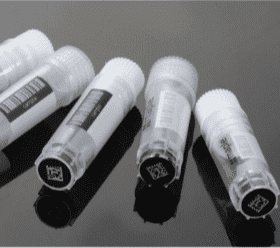
Cryopreservation is a technique designed to preserve the viability and functionality of biological specimens, such as cells, tissues, and organs, by cooling them to subzero temperatures (−80 to −196°C) for long-term storage and transport. By storing biological materials below −130°C, molecular motion is effectively halted, placing cells in a suspended state and significantly slowing biochemical and enzymatic processes, metabolism, and biomolecular transport. This temporary pause in biological activity enables minimal disruption and maintains cellular integrity over extended periods.
As the most reliable method for long-term cell preservation, cryopreservation plays a critical role in cell and gene therapy (CGT) manufacturing. It supports long-term storage, facilitates essential release testing, and enables regulatory documentation and reliable transport under controlled conditions. Advances in cryoshipping technologies now allow for stable temperature maintenance over several days, improving flexibility and overcoming logistical challenges associated with fresh cell shipments.
Despite its advantages, early-stage developers may hesitate to adopt cryopreservation due to concerns about potency loss, added costs, and infrastructure requirements. However, when properly implemented, cryopreservation enhances product stability, supports quality control, and aids regulatory compliance.
Effective cryopreservation strategies are essential for maintaining both starting material integrity and final product quality. Key factors include optimal cooling rates, cryoprotectant composition, and consistent storage conditions to preserve cell viability, functionality, and potency post-thaw. While research labs may rely on non-standardized “home-brew” methods, commercial CGT manufacturing demands GMP-compliant cryopreservation media to ensure reproducibility and regulatory alignment.
By integrating optimized cryopreservation practices into their workflows, CGT developers can improve manufacturing robustness, mitigating risks, and scaling up cell-based therapies with greater confidence and control.
Biological Impact of Cryopreservation
Cryopreservation not only causes physical damage like cell dehydration and osmotic stress but also leads to biological changes, including morphological alterations, protein denaturation, and metabolic and genetic modifications.
Morphological Alterations
Dehydration during freezing results in morphological changes due to altered membrane properties. The “minimum cell volume” model suggests that cells undergo irreversible permeability changes when compressed to their minimum volume.
Effects of dehydration during freezing include:
Changes in membrane properties during dehydration
Lyotropic phase transition in membranes, contributing to ice formation
Actin filament depolymerization and cytoskeleton changes to stabilize the membrane against mechanical stress
Disruption in cell division, especially in embryonic cells
Protein Denaturation
Protein denaturation during freezing involves alterations in protein structure, such as the transition from α-helix to β-sheet.
Cold stress causes native proteins to unfold, exposing nonpolar groups to water.
Though cold-induced denaturation can be reversible under certain conditions, ice formation and changes in pH and electrolyte concentration further disrupt protein structure and activity.
To minimize this, optimizing buffer composition and cooling rates is essential.
Metabolic, Apoptotic, and Genetic Changes
Reactive oxygen species (ROS) increase during cryopreservation, causing damage to proteins, lipids, and DNA
ROS trigger apoptotic pathways and cytochrome c release, leading to cell death
Membrane damage and mitochondrial dysfunction are observed during freezing
DNA double-strand breaks may form due to histone modifications (e.g., H2AX autophosphorylation)
While fetal bovine serum (FBS) helps reduce oxidative stress, its effectiveness decreases when transferrin is saturated with iron.
Why Cryopreservation Matters in Cell Therapy: From Leukopaks to Final Product
Cryopreservation plays a pivotal role in overcoming logistical, operational, and clinical challenges across the entire cell therapy workflow, starting from donor/patient leukopak collection to final drug product delivery. While fresh materials may offer benefits in certain early-phase settings, cryopreservation is essential for scalability, quality control, and commercial readiness.
Logistical Challenges with Fresh Materials
Fresh Leukopaks (Starting Material)
Scheduling Complexity: Aligning donor or patient leukapheresis with immediate downstream processing is logistically demanding. Unpredictable delays—due to illness, lab capacity, or transport issues—can jeopardize material quality.
Time Sensitivity: Fresh leukopaks must typically be processed within 24–36 hours. This short window limits manufacturing flexibility and is only feasible when collection and processing facilities are nearby.
Fresh Final Cell Products
Narrow Delivery Window: Final cell therapies administered fresh have a limited shelf life (often ≤48 hours). Infusion must be tightly coordinated with release testing and clinical site availability.
Transport Risks: Any delay or temperature excursion during shipment can compromise product viability, resulting in missed treatment windows or product wastage.
Advantages of Cryopreservation
For Leukopaks
Enhanced Flexibility: Cryopreserved leukopaks decouple donor/patient collection from manufacturing, enabling asynchronous scheduling and better facility utilization.
Improved Quality Control: Cryopreservation allows sufficient time for sterility, mycoplasma, and identity testing before use in GMP manufacturing.
Inventory Readiness: Enables creation of donor pools and stored materials for platform development or repeated manufacturing runs.
For Final Cell Therapy Products
Reliable Dosing and Redosing: Cryopreserved final products can be stored and thawed on demand, supporting predictable administration schedules, including multiple dosing regimens.
Global Scalability: Frozen drug products can be manufactured centrally and shipped worldwide, supporting multicenter trials and commercial distribution.
Regulatory and QC Efficiency: Enables full product release (including lot release testing) prior to infusion, ensuring only qualified material reaches patients.
Key Benefits of Cryopreservation in Cell Therapy
Benefit
Leukopaks (Starting Material)
Final Cell Products
Scheduling Flexibility
Decouples donor collection from processing
Decouples manufacturing from patient infusion
Transportation Stability
Enables long-distance shipping to central GMP sites
Allows global distribution to clinical centers
Quality Control Buffer
Time for sterility and identity testing pre-manufacture
Enables full release testing before use
Scalability
Supports donor inventory and batch manufacturing
Enables large-scale deployment across sites
Support for Redosing
Stored material can be used for repeat manufacturing
Frozen product can be thawed for scheduled re-dosing
Fresh vs. Cryopreserved Leukopaks: Key Considerations in Cell Therapy Manufacturing
Leukopaks—leukapheresis-derived collections of peripheral blood mononuclear cells (PBMCs)—serve as the foundational starting material in the manufacturing of cell therapies. Whether for autologous or allogeneic products, the choice between fresh and cryopreserved leukopaks can significantly impact process design, product consistency, logistics, and scalability.
Fresh Leukopaks in Cell Therapy
Fresh leukopaks are collected from donors or patients and shipped under ambient or temperature-controlled conditions without cryopreservation. They are especially valuable in autologous settings, where time is critical and minimizing manipulation is preferred.
Represent patient-derived material for autologous T cell or NK cell therapies.
Typically maintain >90% viability for 24–36 hours post-collection; degradation starts beyond this window due to neutrophil degranulation and metabolic stress.
Autologous plasma supplementation may extend the viable use up to 72 hours.
Must be processed promptly upon arrival for T cell isolation, activation, gene editing, and expansion steps.
Logistics for Fresh Leukopaks
Requires same-day or next-day delivery to the manufacturing facility.
Transportation windows must be carefully planned to avoid after-hours or weekend deliveries.
Digital chain-of-custody platforms are essential to ensure traceability and sample integrity.
Advantages for Cell Therapy
High functional integrity of T cells and other immune subsets critical for downstream activation and expansion.
Preferred for autologous manufacturing, where the patient’s own cells are collected, modified, and returned.
Faster vein-to-vein time, a key metric in autologous therapies with aggressive timelines.
No DMSO carryover, eliminating the need for washing steps that can affect yields.
Challenges for Cell Therapy
Logistics-dependent: Fresh leukopaks must be processed within tight timelines (typically 24–36 hours), requiring proximity between collection and manufacturing sites.
Scheduling complexity: leukapheresis, transportation, and GMP processing must be tightly coordinated.
Limited scalability: not feasible for batch manufacturing or large-scale operations due to timing constraints.
Greater risk of product variability due to differences in donor/patient condition and timing.
Cryopreserved Leukopaks in Cell Therapy
Cryopreserved leukopaks are frozen shortly after collection using controlled-rate freezing protocols and stored at ultra-low temperatures (≤ –150°C). They support both autologous and allogeneic workflows, particularly where long-term storage, global shipment, and batch processing are needed.
Preserved using cryoprotectants (typically 5–10% DMSO) to minimize ice crystal damage.
Enable long-term storage, often months to years, while maintaining the functionality of T cells and other PBMCs.
Common in allogeneic “off-the-shelf” cell therapy platforms, where pooled donor cells are used across multiple patients.
Advantages for Cell Therapy
Decouples leukapheresis from manufacturing, providing scheduling flexibility and supporting centralized manufacturing models.
Enables batch testing and pooling strategies, improving process control and scalability.
Allows global collection and centralized GMP processing, reducing time and cost barriers in multicenter trials or commercial operations.
Supports in-process testing and release assays before initiating manufacturing.
Thawing must be tightly controlled to preserve viability and reduce cryo-injury.
Risk of post-thaw viability loss or shifts in cell phenotype if cryopreservation conditions are not optimized.
DMSO toxicity may necessitate additional washing or dilution steps post-thaw.
Some cell surface markers or cytokine responsiveness may be altered after freeze/thaw, impacting activation or gene editing efficiency.
Considerations for Cryopreservation for Cell Therapy
Pre-freeze Processing Activities to Optimize Cell Harvesting Before Cryopreservation
Cells should be harvested during the exponential growth phase, just before entering the stationary phase, to maximize viability and uniformity.
The required cell concentration for cryopreservation varies by cell type and application. Cell biobanking typically use 10⁶ to 10⁷ cells per mL, but other applications may differ.
Renewing the complete growth medium one day before harvest can improve cell health and viability.
Before freezing, cell suspensions should be washed by centrifugation and resuspended in an isotonic medium at a defined concentration. Stressed cells experience higher losses during freezing and reduced post-thaw viability.
Actively growing cells in the exponential phase have a lower cytoplasm-to-nuclear volume ratio, improving cryoprotectant effectiveness. Using standardized culture media and reagents across experiments enhances reproducibility.
The prefreeze phase may involve selecting specific subpopulations of cells, expanding them ex vivo, or incubating them with activating or priming factors to optimize them for cryopreservation.
Cells harvested with plasma should be processed with an anticoagulant to prevent clotting.
Some cells aggregate during centrifugation or are more fragile, necessitating optimized wash steps specific to cell type, suspension volume, and container.
Ideally, cell samples should be tested for adventitious agents before freezing. The testing regimen depends on donor source, culture history, and intended use. Maintaining detailed records supports risk assessment and supplementary testing, such as screening for bovine viruses if bovine serum albumin was used. Regulatory requirements vary based on intended use and past experience in cell therapy and vaccine manufacturing.
Documenting the cells’ pre-cryopreservation characteristics and identity early in the process development is crucial to ensuring consistency and traceability.
Use of Cryoprotectant Solution
Cryopreservation media typically include isotonic saline-based solutions with intracellular cryoprotectants like 5%–10% DMSO and, in some cases, extracellular agents such as hydroxyethyl starch.
The residual DMSO content in the final product should be assessed or quantified.
According to ICH guidelines, DMSO is classified as a Class 3 solvent, indicating relatively low risk in pharmaceutical formulations.
In clinical cell therapy, DMSO exposure is typically limited to 1 g/kg of recipient weight per day.
DMSO has been reported to cause adverse effects in patients, including gastrointestinal, cardiovascular, and neurological toxicities.
To minimize DMSO-related toxicity, premedication with diphenhydramine or other antihistamines is a common practice. Additionally, product washing—whether through manual centrifugation or automated methods—can help reduce residual DMSO levels, but these methods must be validated to ensure sufficient cell recovery and functionality post-wash.
DMSO-free cryopreservation media
DMSO-free cryopreservation media offer several advantages over traditional DMSO-based agents, particularly in sensitive cell types and therapeutic applications:
Enhanced Safety for Sensitive Cells: safer alternatives for cells such as mesenchymal stem cells, induced pluripotent stem cells (iPSCs), primary cells, immune cells, as well as rare and reproductive cells that are particularly vulnerable to DMSO-related toxicity.
Better suited for rare and reproductive cells sensitive to DMSO toxicity.
Reduced Unwanted Differentiation: Avoids DMSO-induced differentiation, preserving cell functionality, which is especially important in research and clinical settings where preserving specific cell functions is critical.
Improved Post-Thaw Viability and Functionality
support better survival rates and retention of potency, even in short-term storage at -80°C, compared to traditional liquid nitrogen storage with DMSO.
These characteristics make them highly suitable for therapeutic use, where consistent cell quality and function are essential.
Therapeutic and Manufacturing Advantages
Safer for direct injection (e.g., into the brain or eye) as dilution is limited.
Facilitates scale-up with more flexible fill/finish processes.
Eliminates the need for wash steps, reducing cost and complexity.
Ensures consistency and reliability across manufacturing batches.
Cooling
Because cryoprotectants like 10% DMSO are hypertonic, they can induce osmotic stress, leading to rapid cell volume changes and reduced viability.
To mitigate this, the cryoprotectant solution should be added gradually—either in stepwise increments or by slow dispensing along the container wall—to prevent excessive cell loss.
Prechilling the medium and cell suspension before adding cryoprotectant can further reduce heat-related cellular stress.
After cryoprotectant addition, the suspension can be transferred to a controlled-rate freezer, generating a freeze curve recorded for production documentation. Temperature probes track product temperature to ensure controlled freezing.
Controlled-rate freezing adjusts nitrogen gas flow to regulate temperature, optimizing cooling protocols for post-thaw recovery. Probes should monitor temperature, and backup plans should be in place for cryogen supply failures.
Passive freezing involves placing samples in a −80°C or −150°C freezer. Insulated containers moderate cooling rates. While controlled freezing offers better consistency, some cells exhibit comparable recovery with passive freezing if parameters are validated.
Cryogenic Storage, Safety, and Transport
Most cell biobanks require liquid nitrogen storage. Insulated transport devices prevent warming during transfer. Newly frozen cells remain in quarantine until tested for adventitious agents.
To prevent temperature fluctuations, infrequently accessed cell biobanks should be stored separately from frequently retrieved cultures. Multiple storage locations reduce catastrophic loss risks.
Liquid nitrogen vapor-phase storage is preferred for safety and ease of retrieval, but routine monitoring is required due to temperature variations.
Cryopreserved cells are shipped in liquid nitrogen vapor shippers with temperature tracking. Shipping containers should be validated under worst-case conditions. Regulatory compliance with IATA and DOT guidelines is essential.
Cells should be thawed rapidly to prevent ice recrystallization and osmotic stress. Optimal thawing procedures should be established for each cell type.
Water baths are commonly used, but therapeutic cell preparations may be thawed in bead baths or thermoblocks to reduce contamination risks. Water baths should be regularly cleaned and monitored for consistency.
Thawing rates should exceed 1°C per second. While temperatures above 42°C can accelerate thawing, excessive heat may induce necrosis or apoptosis. Careful validation is needed to balance efficiency and viability.
Clinical sites handling cryopreserved products should have properly trained personnel, appropriate storage facilities, and validated thawing procedures to ensure product integrity and patient safety.
Although bedside thawing for administration of cell therapy products is common, controlled laboratory thawing is preferable for standardization and sterility. Thawing is typically performed in a 37°C water bath, with overwrap bags reducing contamination risks. Gentle kneading of the bag ensures uniform thawing.
Post-Thaw Processing and Evaluation
Post-thaw processing is often required since cryopreservation solutions are not physiologically compatible. For DMSO-preserved cells, immediate washing or dilution is necessary to prevent cytotoxicity.
Minimum post-thaw viability thresholds should be established. Any product below these limits should be discarded. Post-thaw assays should be developed and validated to ensure product consistency.
The cryopreservation process must consider cell viability, function, and population shifts, as some cell subtypes are more vulnerable to freeze–thaw damage.
Mechanical activity (attachment or contraction assays)
Mitotic activity (proliferation assays)
Engraftment potential (for therapeutic cells)
Using at least two independent viability methods is recommended. Cryopreserved cell stability should be periodically assessed by thawing representative vials (per ICH Q5D guidelines).
Contamination Testing
Routine testing for adventitious agents is necessary for Master Cell Banks (MCBs) and Working Cell Banks (WCBs). USP general chapters 〈1050〈 and 〈1237〈 provide guidance.
Standard sterility testing may not detect mycobacteria, so additional mycoplasma and bacterial contamination testing should be considered. USP 〈63〈 outlines mycoplasma detection methods.
Periodic screening for bacterial, fungal, mycoplasma, and viral contaminants is recommended.
Documentation and Quality Control
Batch records should document cell history from receipt to cryopreservation, storage, and use. Key details include:
Cryopreservation procedure followed
Equipment used (with unique identifiers)
Freezing profile records
Post-thaw viability assessments
Storage and retrieval logs
Longitudinal monitoring confirms process stability. Documentation should align with quality management systems to ensure traceability and regulatory compliance.
Regulatory Considerations for Cell Therapy Applications
Regulatory Challenges of Cryopreservation
Impact on Cell Quality
Freeze/thaw stress may impair viability, functionality, or phenotype, especially in sensitive cell types like NK cells or γδ T cells.
Cryopreservation can affect membrane integrity, metabolic activity, or cytotoxic function.
Variability in freeze rate, thawing method, and handling conditions can affect consistency.
FDA Expectations for Cryopreserved Cell Therapy Products
According to FDA guidance, the cryopreservation process must be standardized and validated to ensure it does not compromise critical quality attributes (CQAs). Key expectations include:
GMP-Compliant Cryopreservation Media
Avoidance of animal-origin components unless thoroughly justified.
Reagents and excipients must meet human-use safety and stability criteria.
Control of Cryopreservation-Induced Variability
Pre- and post-thaw comparisons for CQAs (e.g., viability, identity, potency).
Demonstrated equivalence between fresh and cryopreserved product, if applicable.
Final Formulation and Thawing
FDA prefers infusion-ready formulations to reduce manipulation at clinical sites.
Justification is needed for any post-thaw processing steps, like washing or dilution.
Stability Analysis of Cryopreserved Cell Therapy Products
Stability studies are a critical component of the Chemistry, Manufacturing, and Controls (CMC) section for IND and BLA submissions. These studies assure that the product maintains its identity, strength, quality, and purity throughout its intended shelf life.
Key Aspects of Stability Testing
Real-Time and Accelerated Conditions: Conducted under actual and stressed storage conditions (e.g., −150°C, −80°C, dry ice) to evaluate degradation patterns and inform labeling claims.
Potency assays: Must be quantitative and stability-indicating (e.g., IFN-γ ELISPOT, target cell lysis).
Stability-Indicating Assays: Potency assays must be sensitive to degradation over time and validated for specificity, accuracy, and precision.
Establishing Shelf Life: Initial shelf-life is based on real-time data, but accelerated studies may support early clinical use. Long-term studies must continue throughout development.
Documentation Required in IND/BLA:
Stability protocol with time points and testing rationale
Description of sample handling and thawing conditions
Acceptance criteria and justification for specification limits
Data summaries showing that the product remains within specifications during shelf life
Cryopreservation is not merely a logistical enabler—it is a cornerstone of quality assurance and regulatory compliance in cell therapy manufacturing. From leukopak starting materials to the final cell therapy product, a well-defined and validated cryopreservation strategy is essential to maintaining product integrity, therapeutic potency, and patient safety across the supply chain.
Successfully navigating the regulatory landscape requires demonstrating that freezing and thawing processes do not adversely impact critical quality attributes. This includes establishing GMP-grade cryopreservation media and protocols, validating stability-indicating assays, ensuring post-thaw viability and potency, and clearly defining shelf life based on robust stability data.
As cell therapies advance from early-phase trials to global commercialization, cryopreservation offers the scalability, flexibility, and control necessary for consistent delivery of high-quality products. A comprehensive approach—rooted in scientific rigor and regulatory alignment—ensures that the promise of cell therapy is preserved from collection to clinic.
References
Bai et al. (2023) Cryopreservation in the Era of Cell Therapy: Revisiting Fundamental Concepts to Enable Future Technologies. Advanced Functional Materials. Volume 33, Issue 40
Seaver S. A new United States Pharmacopeia (USP) Chapter 1046: cell and gene therapy products. Cytotherapy. 2000;
FDA Guidance for Industry: Considerations for the Development of CAR T Cell Products (Jan 2024)
FDA Guidance for Industry: Chemistry, Manufacturing, and Control (CMC) Information for Human Gene Therapy Investigational New Drug Applications (INDs) (Jan 2020)
The concept of Critical Quality Attributes (CQAs) plays a pivotal role in a drug development process. According to the ICH Guideline Q8(R2) on Pharmaceutical Development,
Cell and gene therapies (CGTs), particularly autologous therapies like CAR-T/TCR-T cells, represent a breakthrough in personalized medicine. These treatments offer life-changing outcomes for patients with
Extracellular vesicles (EVs) have emerged as critical players in diagnostics, vaccine development, and therapeutic applications, driving the need for precise characterization tools. Exoid, powered by
HOW CAN WE HELP YOU?Our specialists are to help you find the best product for your application. We will be happy to help you find the right product for the job.