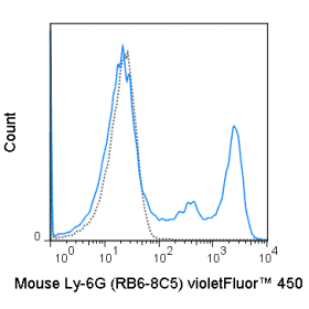
The RA3-6B2 antibody reacts with the human and mouse CD45 isoform known as CD45R, or B220, a protein tyrosine phosphatase of ≥ 220 kDa. CD45 is one of the most abundant hematopoietic markers, and is expressed on all leukocytes (the Leukocyte Common Antigen, LCA). Various isoforms are generated and expressed in cell-specific patterns, all critical for leukocyte function. In mouse, the CD45R/B220 isoform is predominantly found on B cells, at varying levels on all stages from pro-B cells to activated B cells, and may also be detected on certain T cell and NK cell subsets. It is of note that B220 is not similarly expressed on human B cells, where it appears to be differentiation-specific and therefore expressed on only some B cell subsets. Other forms of CD45 with restricted cellular expression include CD45RA, CD45RB, CD45RO and several others.
The RA3-6B2 antibody is one of the most consistently used leukocyte markers for B cells, T cell subsets and NK cell subsets in human and mouse. This antibody has also been reported as cross-reactive with feline CD45R/B220.
Product Details
| Name | PerCP-Cyanine5.5 Anti-Human/Mouse CD45R (B220) (RA3-6B2) |
|---|---|
| Cat. No. | 65-0452 |
| Alternative Names | Ly-5, Lyt-4, T200 |
| Gene ID | 19264 |
| Clone | RA3-6B2 |
| Isotype | Rat IgG2a, κ |
| Reactivity | Human, Mouse |
| Cross Reactivity | Feline |
| Format | PerCP-Cyanine5.5 |
| Application | Flow Cytometry |
| Citations* | Willinger T and Flavell RA. 2012. Proc. Natl. Acad. Sci. 109:8670-8675. (Flow cytometry)
Meredith MM, Liu K, Darrasse-Jeze G, Kamphorst AO, Schreiber HA, Guermonprez P, Idoyaga J, Cheong C, Yao K-H, Niec RE, and Nussenzweig MC. 2012. J. Exp. Med. 209: 1153-1165. (Immunofluorescence microscopy: acetone-fixed frozen tissue) Becker-Herman A, Meyer-Bahlburg A, Schwartz MA, Jackson SW, Hudkins KL, Liu C, Sather BD, Khim S, Liggitt D, Song W, Silverman GJ, Alpers CE and Rawlings DJ. 2011. J. Exp. Med. 208:2033-2042. (Immunofluorescence microscopy – OCT embedded frozen tissue) Bertossi A, Aichinger M, Sansonetti P, Lech , Neff F, Pal M, Wunderlich FT, Anders H, Klein L, and Schmidt-Supprian M. 2011. J. Exp. Med. 208:1749-1756. (Immunofluorescence microscopy) De Clercq S, Gembarska A, Denecker G, Maetens M, Naessens M, Haigh K, Haigh JJ, and Marine J-C. 2010. Mol. Cell. Biol. 30:5394-5405. (Western Blot) Nutt SL, Metcalf D, D’Amico A, Polli M, and Wu L. 2005. J. Exp. Med. 201:221-231. (Immunomagnetic bead depletion) Cappione AJ, Pugh-Bernard AE, Anolik JH, and Sanz I. 2004. J. Immunol. 172: 4298-4307. (Immunoprecipitation) Monteith CE, Chelack BJ, Davis WC, and Haines DM. 1996. Can. J. Vet. Res. 60(3): 193-198. (Immunohistochemistry – feline tissue) Whiteland JL, Nicholls SM, Shimeld C, Easty DL, Williams NA, and Hill TJ. 1995. J. Histochem. Cytochem. 43:313-320. (Immunohistochemistry – frozen tissue, paraffin embedded tissue) Domiati-Saad R, Ogle EW, and Justement LB. 1993. J. Immunol. 151: 5936-5947. (in vivo blocking) |
Application Key:FC = Flow Cytometry; FA = Functional Assays; ELISA = Enzyme-Linked Immunosorbent Assay; ICC = Immunocytochemistry; IF = Immunofluorescence Microscopy; IHC = Immunohistochemistry; IHC-F = Immunohistochemistry, Frozen Tissue; IHC-P = Immunohistochemistry, Paraffin-Embedded Tissue; IP = Immunoprecipitation; WB = Western Blot; EM = Electron Microscopy
*Tonbo Biosciences tests all antibodies by flow cytometry. Citations are provided as a resource for additional applications that have not been validated by Tonbo Biosciences. Please choose the appropriate format for each application and consult the Materials and Methods section for additional details about the use of any product in these publications.


